Fossil Taxonomy – Reduviasporonites


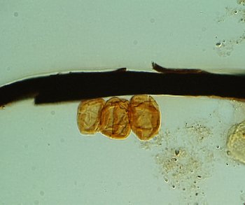
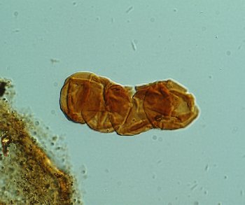
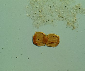
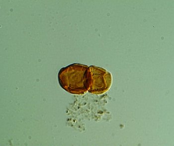
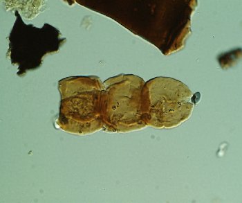
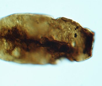
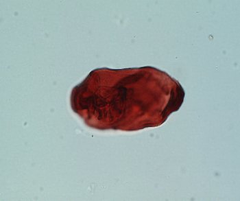
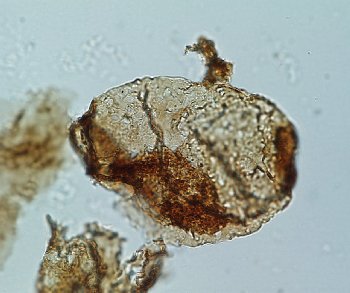
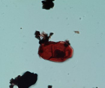
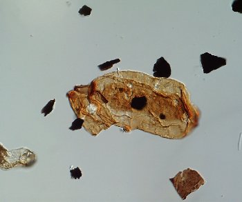
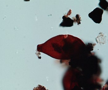
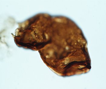
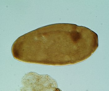
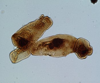
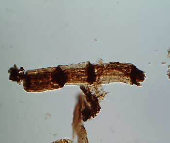
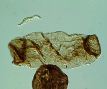
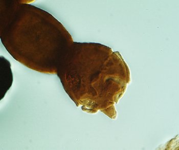
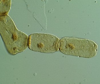
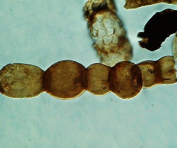
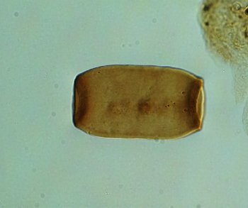
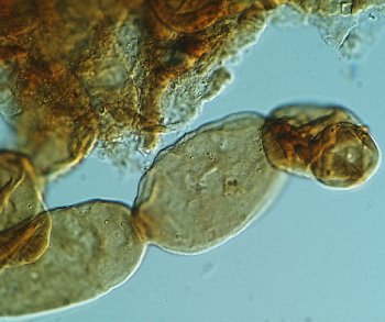
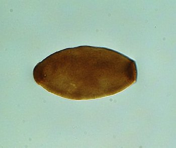
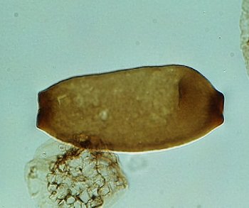
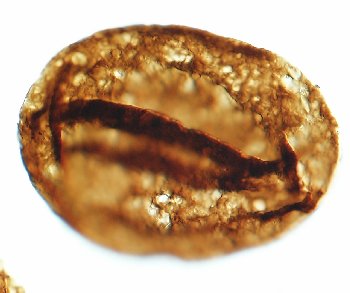
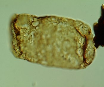
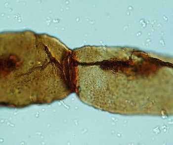
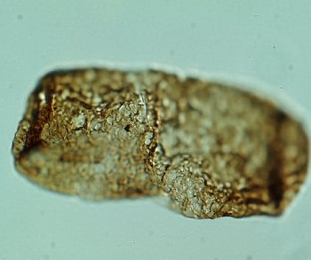
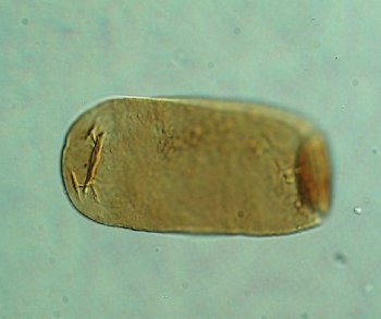
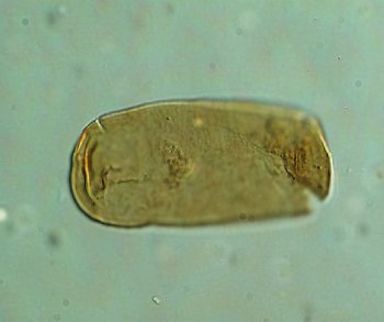
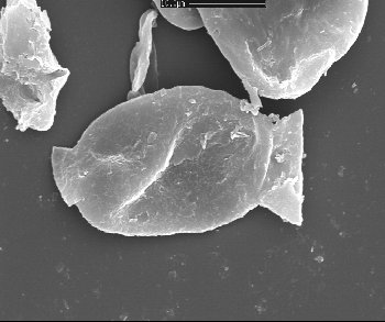
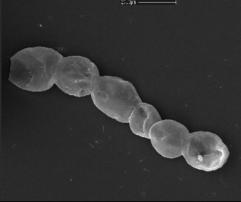
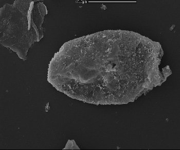
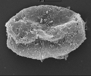
Reduviasporonites catenulatus Wilson 1962
Chain of 5 cells; left outer cell without a terminal thickening or rim.
Mean length of cells 15µm
USA
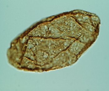
C34
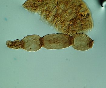
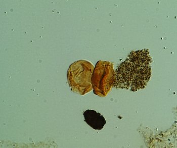
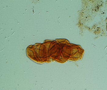
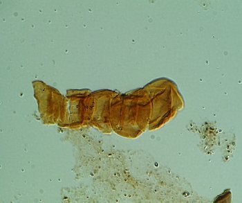
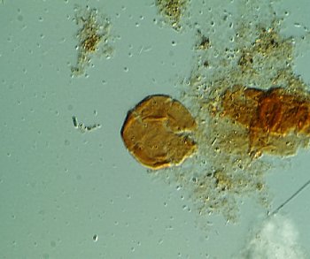
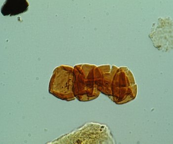
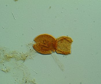
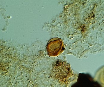
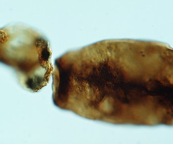
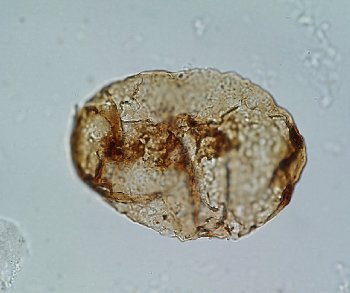
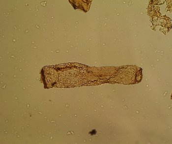
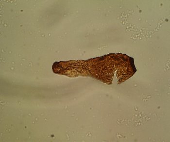
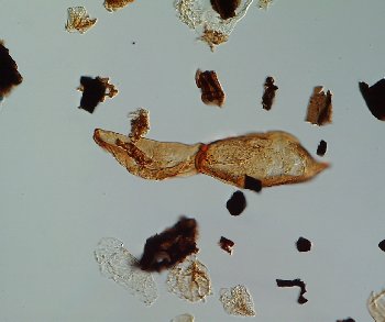
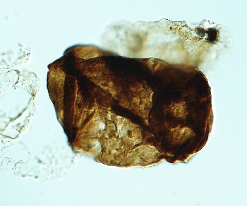
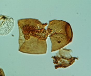
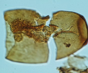
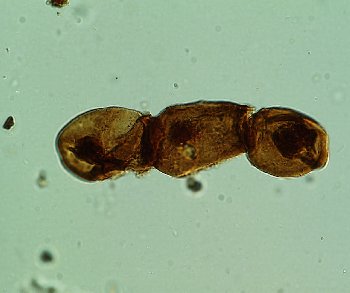
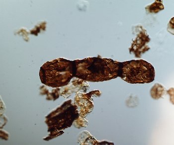
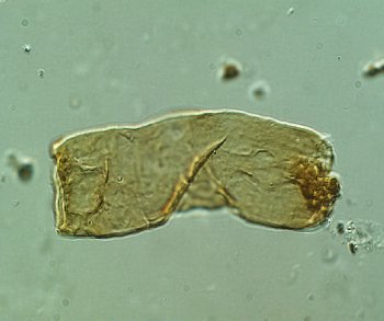
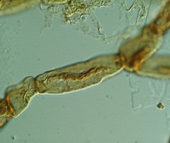
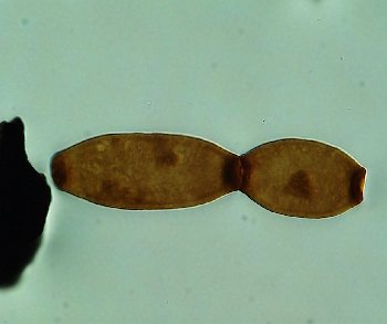
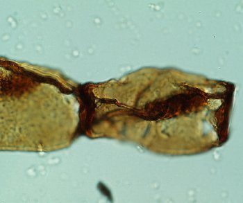
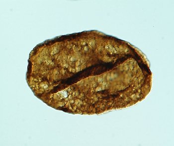
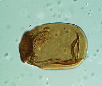
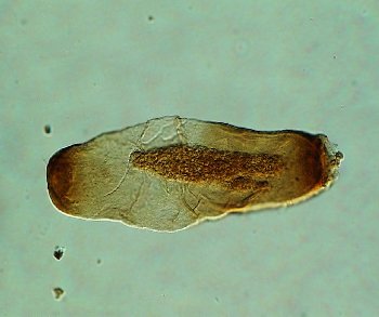
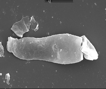
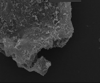
Reduviasporonites catenulatus Wilson 1962
Poorly preserved ?double chain
Mean length of cells 15µm
USA
D43
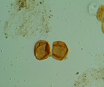
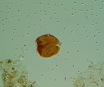
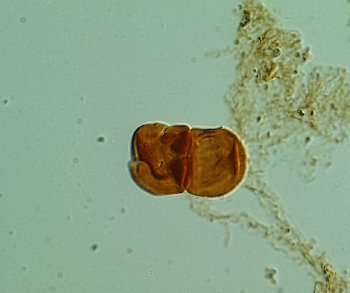
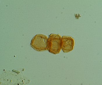
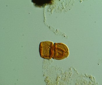
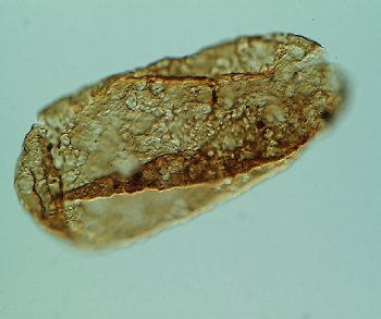
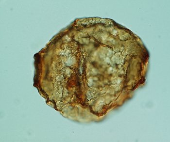
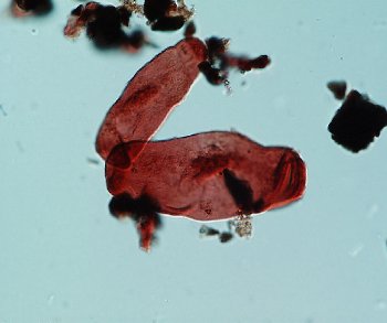
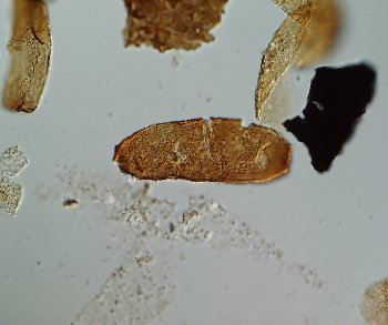
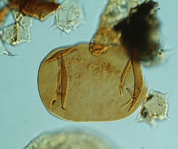
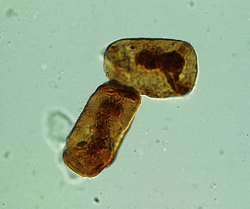
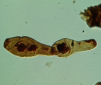
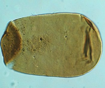
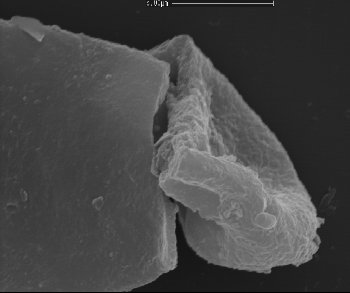
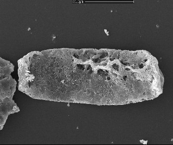
Reduviasporonites catenulatus Wilson 1962
Two cells in chain.
Mean length of cells 11µm
USA
E15
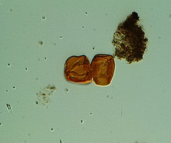
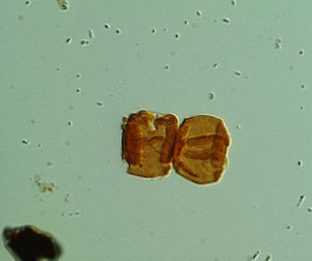
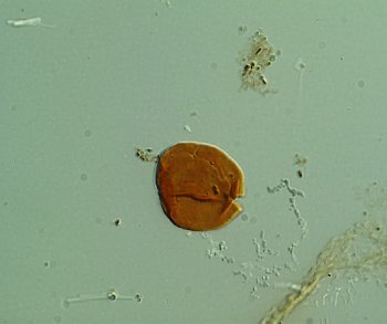
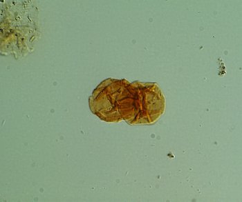
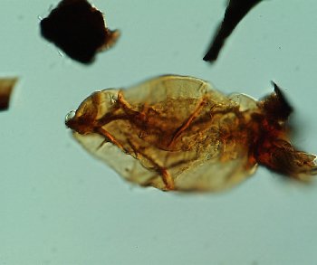
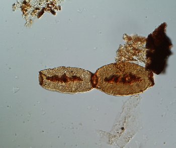
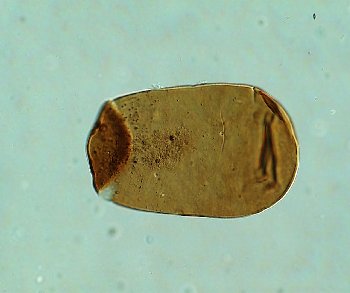
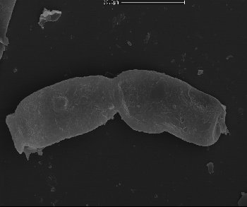
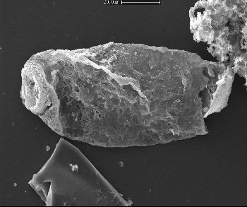
Reduviasporonites catenulatus Wilson 1962
Two cells in chain.
Mean length of cells 12µm
USA
E18
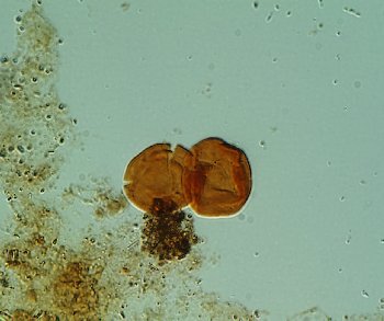
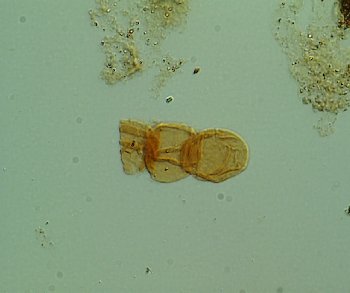
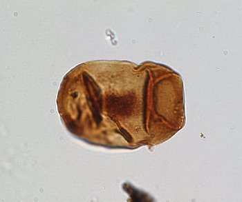
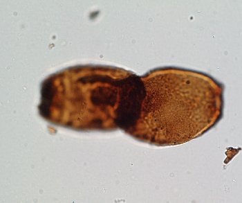
Reduviasporonites catenulatus Wilson 1962
Two recently separated cells.
Mean length of cells 12µm
USA
E19
Reduviasporonites catenulatus Wilson 1962
Two cells in chain.
Mean length of cells 14µm
USA
E19
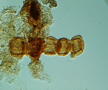
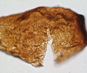
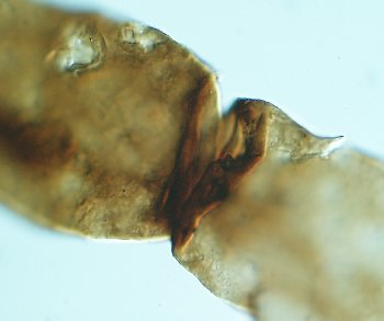
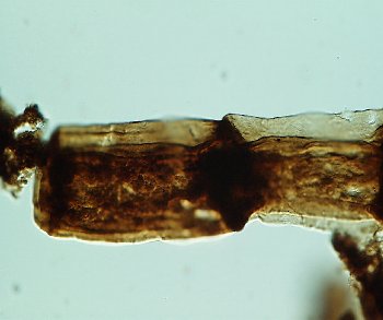
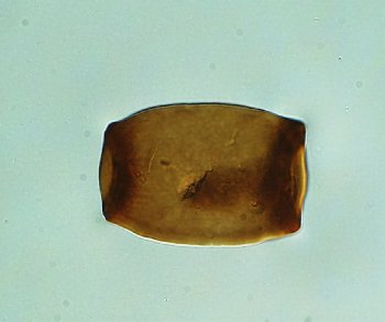
Reduviasporonites catenulatus Wilson 1962
Chain of cells; neither has an outer terminal rim or thickening. Left cell shows inner body through the crack in the outer cell wall.
Mean length of cells 16µm
USA
E19/4
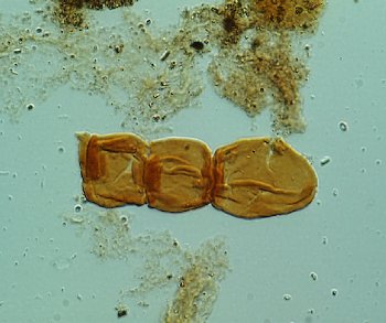
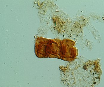
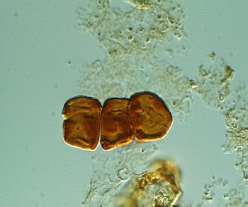
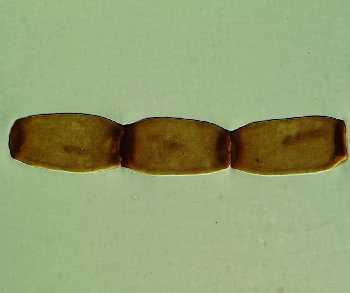
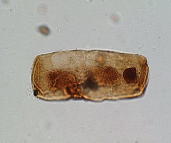
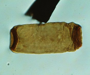
Reduviasporonites catenulatus Wilson 1962
Chain of three cells; neither of the outer two cells has an outer terminal rim or thickening.
Mean length of cells 11µm
USA
E23
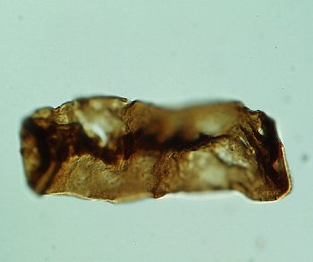
Reduviasporonites catenulatus Wilson 1962
Chain with longitudinally collapsed cells.
Mean length of cells 15µm
USA
E36/4
Reduviasporonites catenulatus Wilson 1962
Chain with longitudinally collapsed cells.
Mean length of cells 14µm
USA
G16
Reduviasporonites catenulatus Wilson 1962
Chain with relatively well preserved cells close in form to some of the Alpine specimens. The right edge of the right hand cell appears not to have been connected to another cell. The left cell has clearly been detached from another cell.
Lengths of cells 12, 13 and 22µm.
USA
G36
Reduviasporonites catenulatus Wilson 1962
Chain with longitudinally collapsed cells.
Mean length of cells 14µm
USA
G39/2
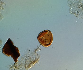
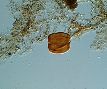
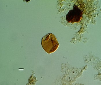
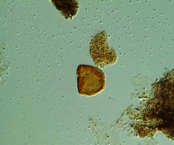
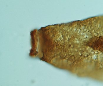
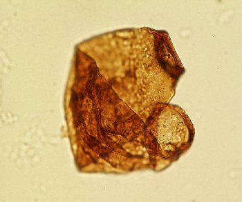
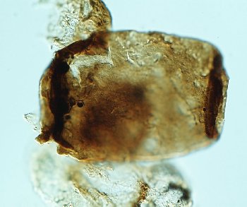
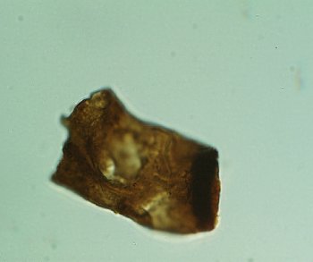
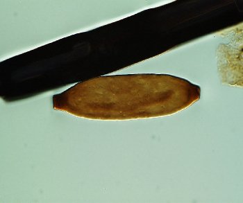
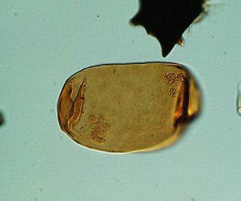
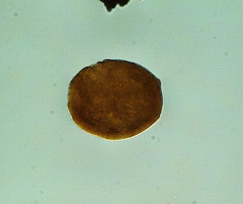
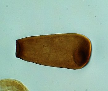
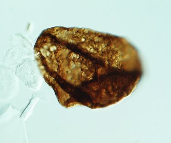
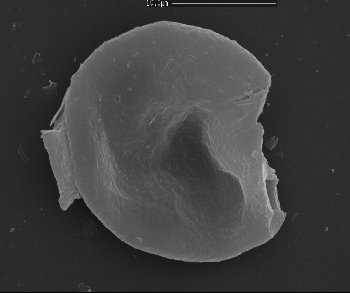
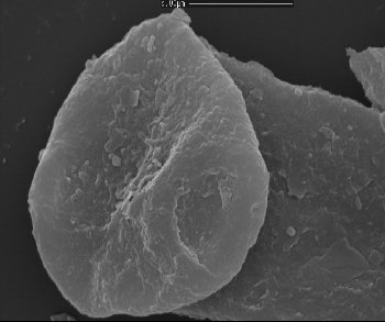
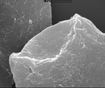
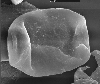
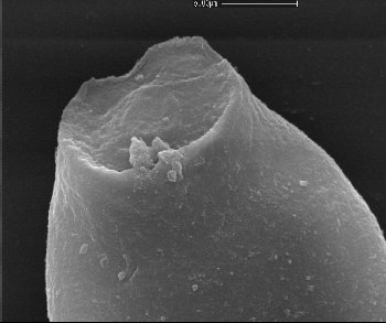
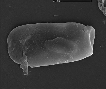
Reduviasporonites catenulatus Wilson 1962
Single cell; perhaps an ‘end cell’. Right side of cell has no terminal rim or thickening. Left side with thickening, possibly was part of a chain in which it was the ‘end cell’.
Cell length 20µm
USA
G41
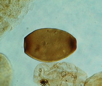
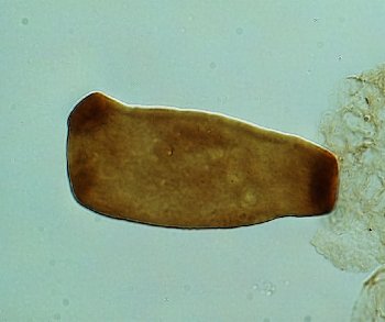
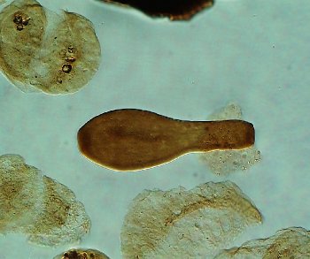
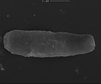
Reduviasporonites catenulatus Wilson 1962
Single cell
Cell length 19µm
USA
G44
Reduviasporonites catenulatus Wilson 1962
Chain with relatively well preserved cells close in form to some of the specimens from the Mazzin Member, Austria. The right edge of the right hand cell appears not to have been connected to another cell. The left cell has clearly been detached from another cell.
Mean length of cells 12µm
USA
H10/2
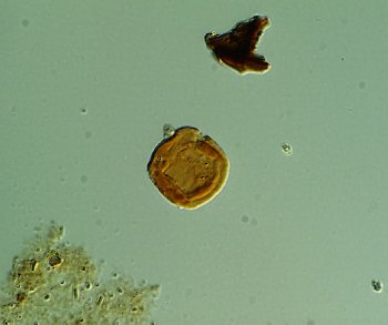
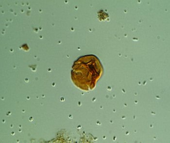
Reduviasporonites catenulatus Wilson 1962
Single cell with a discontinuous concentric pattern of folds
Approx. length 15 µm
USA
H40/4
Reduviasporonites catenulatus Wilson 1962
Chain with longitudinally collapsed cells.
Mean length of cells 13µm
USA
J11/2
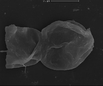
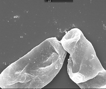
Reduviasporonites catenulatus Wilson 1962
A pair of joined cells. Neither of the outer cells appears to have been connected to other cells.
Mean length of cells 13µm
USA
J24
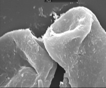
Reduviasporonites catenulatus Wilson 1962
A pair of joined cells with considerable overlap. Neither of the outer cells appears to have been connected to other cells.
Mean length of cells 16µm
USA
J27/3
Reduviasporonites catenulatus Wilson 1962
Chain with longitudinally collapsed cells. Neither of the outer cells appears to have been connected to other cells.
Mean length of cells 15µm
USA
J32
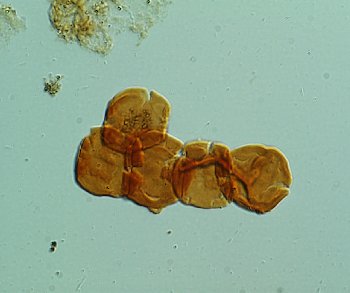
Reduviasporonites catenulatus Wilson 1962
Chain of 4 longitudinally collapsed cells. Left cell displays strong concentric fold pattern, perhaps indicating a folded inner body or perhaps a system of invaginations in the outer cell wall.
Mean length of cells 13µm
USA
L24/4
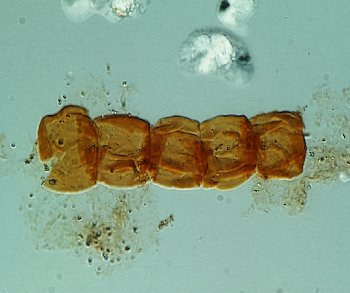
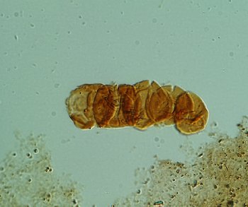
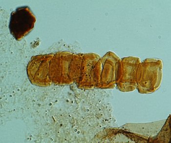
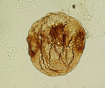
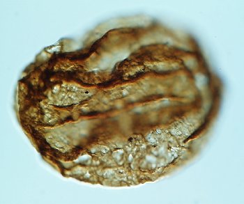
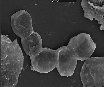
Reduviasporonites catenulatus Wilson 1962
Chain of 5 cells each with a concentric to rectilinear pattern of folds.
Mean length of cells 13µm
USA
L27/4
Reduviasporonites catenulatus Wilson 1962
Chain of three rigid cells. Neither of the outer cells appears to have been connected to other cells.
Mean length of cells 14µm
USA
M24/1
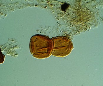
Reduviasporonites catenulatus Wilson 1962
A pair of joined cells.
Mean length of cells 12µm
USA
M29/2
Reduviasporonites catenulatus Wilson 1962
A pair of joined cells. The left hand cell does not appear to have been connected to other cells.
Mean length of cells 18µm
USA
M39/1
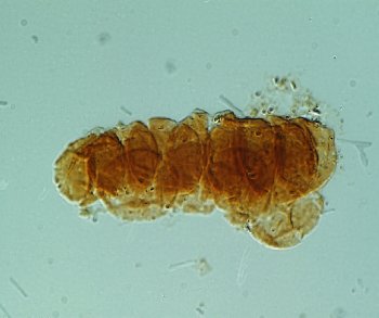
Reduviasporonites catenulatus Wilson 1962
Chain of 6 cells. Most cells display strong concentric fold pattern. There appears to be continuity across the folding patterns of the cells suggesting a possible series of connected inner bodies.
Mean length of cells 13µm
USA
N23/4
Reduviasporonites catenulatus Wilson 1962
Chain of 3 cells. All cells display strong concentric fold pattern.
Lengths of cells 11, 13 and 14µm
USA
N25
Reduviasporonites catenulatus Wilson 1962
Pair of cells. Left hand cell has an invaginated or thickened left margin possible suggesting connection with other cells. Right cell seems to lack means of connection.
Mean length of cells 13µm
USA
N34/4
Reduviasporonites catenulatus Wilson 1962
Chain of 4 cells and a single off line cell possibly the remaining cell of a branch of cells. Cells distinctly overlap each other.
Mean length of cells 12µm
USA
P32
Reduviasporonites catenulatus Wilson 1962
Single cell with a concentric to rectilinear pattern of folds.
Approx. length 15 µm
USA
S15
Reduviasporonites catenulatus Wilson 1962
Single cell with a concentric to rectilinear pattern of folds.
Approx. length 15 µm
USA
S33
Reduviasporonites catenulatus Wilson 1962
Single cell with a concentric pattern of folds.
Approx. length 15 µm
USA
S35
Reduviasporonites catenulatus Wilson 1962
Barrel shaped cell. May have been part of a chain. Has two possible outer invaginations which may indicate site of two previous connections.
Approx. length 15 µm
USA
T12/2
Reduviasporonites catenulatus Wilson 1962
A pair of joined cells. Both cells appear to lack an outward facing joining structure. These were probably never part of a larger chain.
Mean length of cells 13µm
USA
T20
Reduviasporonites catenulatus Wilson 1962
Single cell with fold or invagination at the right margin.
Approx. length 15 µm
USA
T22/1
Reduviasporonites catenulatus Wilson 1962
A pair of joined cells. Both cells appear to lack an outward facing joining structure. These were probably never part of a larger chain.
Mean length of cells 13µm
USA
T36
Reduviasporonites catenulatus Wilson 1962
Single cell irregularly arranged fold
Approx. length 15 µm
USA
T7/1
Reduviasporonites catenulatus Wilson 1962
Single cell with a concentric to rectilinear pattern of folds.
Approx. length 15 µm
USA
U14
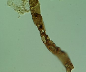
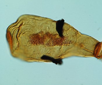
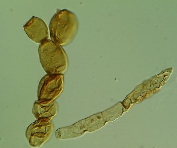
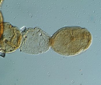
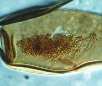
Reduviasporonites catenulatus Wilson 1962
Chain of ovoid cells
Length of cells 15, 20 and 17 µm
USA
Y41/1
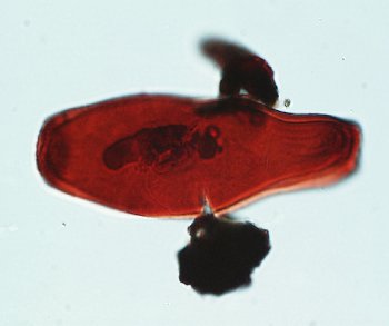
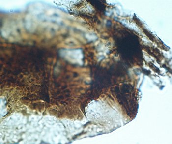
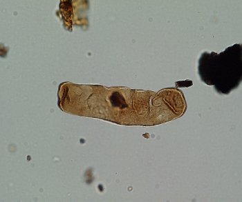
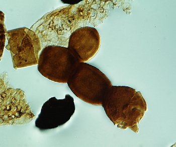
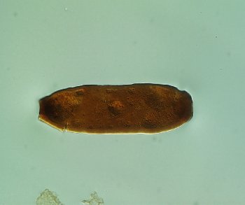
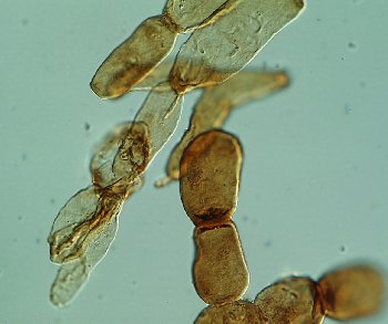
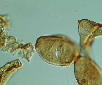
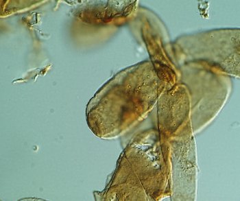
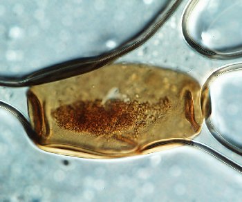
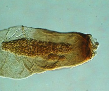
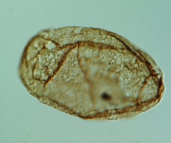
Reduviasporonites catenulatus Wilson 1962
Detail of specimen shown in image Flowerpt 66398_Y41_1. Chain of ovoid cells. The cells to the right both possibly contain slightly shrunken inner bodies. In the middle cell the inner body is attached to the inner surface of the terminal rims on both sides of the cell. Terminal rims are not well displayed between the two right hand cells. Both the inner body wall and the outer cell wall appear to be smooth and less than 1µm thick.
Length of cells 15, 20 and 17 µm
USA
Y41/1
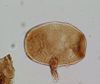
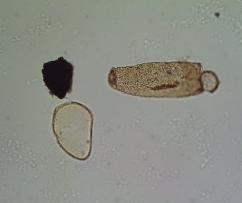
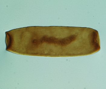
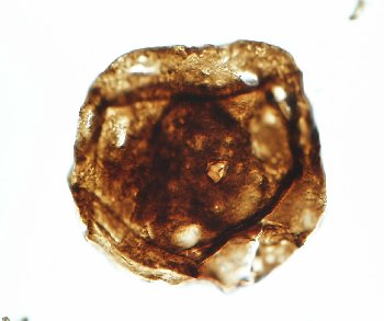
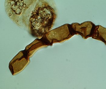
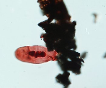
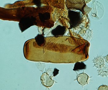
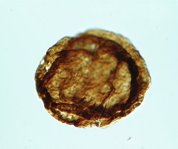
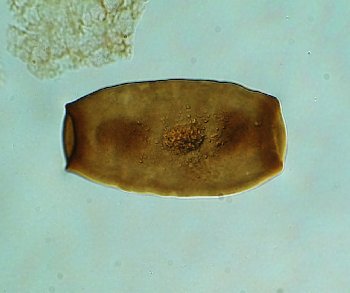
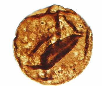
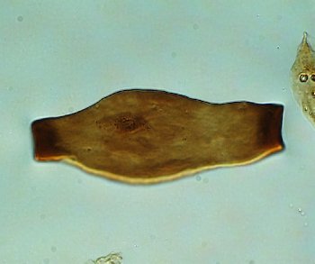
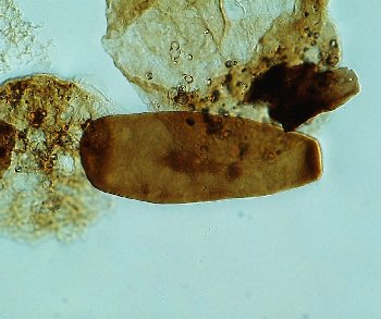
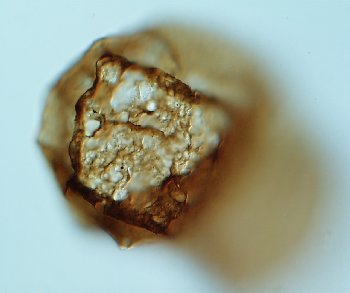
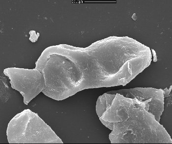
Reduviasporonites chalastus (Foster) Elsik 1999
Elongate cell bent in the centre. Right hand part of the cell shows ‘striations’ in the outer cell wall orientated parallel to the long axis of the cell. These striations may be the result of shrinkage of the outer cell wall.
Length of cell100µm approx.
UK
A27
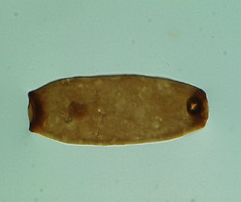
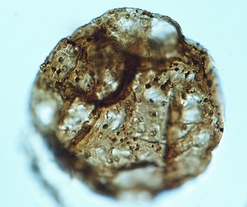
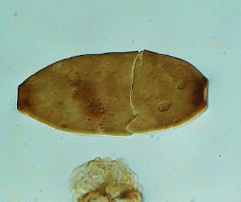
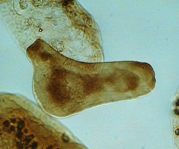
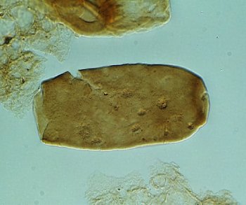
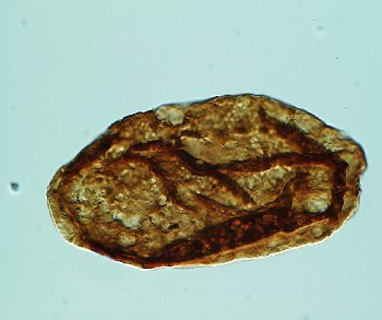
Reduviasporonites chalastus (Foster) Elsik 1999
Elongate cell. Outer cell surface is very finely, indistinctly granulate. Grana less than 0.5µm high and wide; similar distance apart. Inner cell material distinct; amorphous to granular in appearance.
Length of cell 82µm
UK
A28
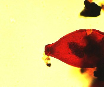
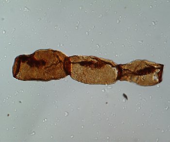
Reduviasporonites chalastus (Foster) Elsik 1999
Well preserved cell with shrunken inner body. Distinct terminal rims.
Length of cell 144µm
Greenland
A52/3
Reduviasporonites chalastus (Foster) Elsik 1999
Detail of specimen shown in image MFP12114_3_750_6_A52_3. Detail of inner body. Terminus of internal body detached from the inner surface of the outer wall.
Length of cell 144µm
Greenland
A52/3
Reduviasporonites chalastus (Foster) Elsik 1999
Detail of specimen shown in image MFP12114_3_750_6_A52_3. Detail of terminal rim.
Length of cell 144µm
Greenland
A52/3
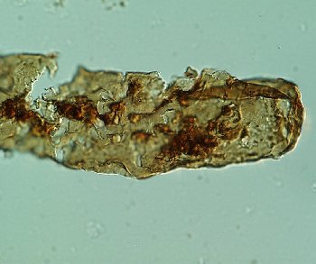
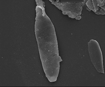
Reduviasporonites chalastus (Foster) Elsik 1999
Long filament with no terminal rims. May not be assignable to Reduviasporonites
Length of cell 190µm approx.
Greenland
B21/1
Reduviasporonites chalastus (Foster) Elsik 1999
Dark coloured (thick walled) specimen; displays very distinct inner and outer cell walls. Outer cell wall 1-1.5µm thick; outer surface very finely granulate (grana only just resolvable under oil x100). Faint shrinkage folds in the outer cell wall are present close to the termini. In these areas the terminal rims are ‘pinched’ i.e. much narrower than usual perhaps called by shrinkage. Inner cell wall 1 µm thick or less; appears smooth. Detached from the inner surface of the outer cell wall only close to the termini. Width of cavity between the two cell walls max. 3µm. Termini of the inner body still attached to the inner surface of the terminal rims. Small cavity at the terminus of the inner body visible on the right side of the cell. Cell material amorphous.
Length of cell 71µm approx.
UK
B27
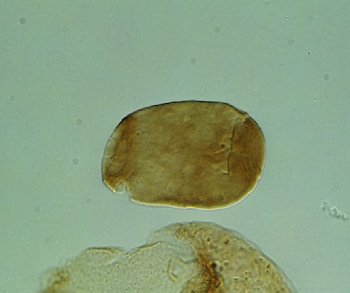
Reduviasporonites chalastus (Foster) Elsik 1999
Small cell with sub-rectangular outline.
Length of cell 40µm
UK
B31/2
Reduviasporonites chalastus (Foster) Elsik 1999
Cell with one indistinct terminal rim
Cell length 53µm
UK
B34/3
Reduviasporonites chalastus (Foster) Elsik 1999
Outer wall of the cell translucent. Inner body distinct; attachment to the inner surface of the outer wall distinct on the left side.
Length of cell 142µm
Greenland
B39/3
Reduviasporonites chalastus (Foster) Elsik 1999
Detail of specimen shown in MFP12114_3_750_6_B39_3. Detail of inner body attachment to the inner part of the terminal rim. Left side
Length of cell 142µm
Greenland
B39/3
Reduviasporonites chalastus (Foster) Elsik 1999
Detail of specimen shown in MFP12114_3_750_6_B39_3. Detail of inner body attachment to the inner part of the terminal rim. Right side
Length of cell 142µm
Greenland
B39/3
Reduviasporonites chalastus (Foster) Elsik 1999
Spindle-shaped cell with dark cell material.
Length of cell 55µm
UK
B47/2

Reduviasporonites chalastus (Foster) Elsik 1999
Well preserved cell with shrunken inner body. Distinct terminal rims. Shows the inner body remains attached to the inner part of the terminal rims.
Length of cell 210µm
Greenland
B9
Reduviasporonites chalastus (Foster) Elsik 1999
Detail of specimen shown in image MFP12114_3_750_6_B9. Detail of well preserved cell with shrunken inner body; showing ‘striations’ in the outer wall.
Length of cell 210µm
Greenland
B9
Reduviasporonites chalastus (Foster) Elsik 1999
Elongate cell with shrunken inner body detached at both ends from the cell termini.
Length of cell 135µm
China
C12
Reduviasporonites chalastus (Foster) Elsik 1999
Ovoid cell
Length of cell 95µm
China
C20/4
Reduviasporonites chalastus (Foster) Elsik 1999
Ovoid cell
Length of cell 95µm
China
C20/4
Reduviasporonites chalastus (Foster) Elsik 1999
Cell with circular outline; terminal rims distinct, inner body shrunken and cell material present. Cf. B. helbyi f. gregata Foster 1979
Length of cell 85µm
China
C20/4
Reduviasporonites chalastus (Foster) Elsik 1999
Terminal rim partially detached suggesting incipient weakness in the outer wall just adjacent to the rim.
Length of cell 125µm
China
C26
Reduviasporonites chalastus (Foster) Elsik 1999
Very small ovoid cell. Cell material amorphous and granular. Very distinct separation between inner body and cell wall. Outer cell wall 1-1.5µm thick; outer surface very finely granulate (grana only just resolvable under oil (x100)). Cell material confined by folds in the inner body.
Length of cell 46µm.
UK
C29
Reduviasporonites chalastus (Foster) Elsik 1999
Elongate cell
Cell length 73µm
UK
C29/3
Reduviasporonites chalastus (Foster) Elsik 1999
Elongate specimen with sub-rectangular outline. Inner body appears adpressed to the inner surface of the cell.
Length of cell 65µm.
Saudi Arabia
C33
Reduviasporonites chalastus (Foster) Elsik 1999
Elongate specimen with sub-rectangular outline. Inner body appears adpressed to the inner surface of the cell.
Length of cell 73µm.
Saudi Arabia
C38
Reduviasporonites chalastus (Foster) Elsik 1999
Elongate cell lacking inner body.
Length of cell 194µm
China
C7/1
Reduviasporonites chalastus (Foster) Elsik 1999
Showing incipient weakness at the base of the terminal rim.
Length of cell 115µm
China
D17/4
Reduviasporonites chalastus (Foster) Elsik 1999
Cell with one indistinct terminal rim and distinct cell material
Cell length 43µm
UK
D30/2
Reduviasporonites chalastus (Foster) Elsik 1999
Chain of three cells which are elongate to ovoid in outline.
Mean length of cells 30µm
Saudi Arabia
D31/1
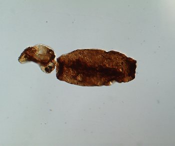
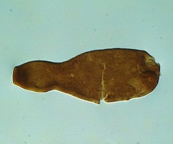
Reduviasporonites chalastus (Foster) Elsik 1999
Flask shaped cell.
Length of cell 160µm
China
D35
Reduviasporonites chalastus (Foster) Elsik 1999
Detail of specimen shown in image 6403380_NLD36_D35. Flask shaped cell. Detail shows that the inner body is missing.
Length of cell 160µm
China
D35
Reduviasporonites chalastus (Foster) Elsik 1999
Cell folded showing terminal rims.
Approx. length of cell 100µm
China
D38
Reduviasporonites chalastus (Foster) Elsik 1999
2 cell chain with short rounded cells
Lengths of cells 57 and 60µm
UK
D38/2
Reduviasporonites chalastus (Foster) Elsik 1999
Very large irregularly shaped cell with distinct inner body.
Length of cell 147µm.
Russia
D41/4
Reduviasporonites chalastus (Foster) Elsik 1999
Two cells, both lacking inner bodies, connected by distinct terminal rims. Significantly the outer ends of both the cells have very poorly developed terminal rims.
Length of cells 105 and 95µm.
Russia
D47
Reduviasporonites chalastus (Foster) Elsik 1999
Ovoid cell with rectilinear folds possibly in the inner body.
Length of cell 130µm
Australia
D51/2
Reduviasporonites chalastus (Foster) Elsik 1999
Cell with irregular outline and a thin outer cell wall (0.5µm or less); contains a distinct, highly folded inner body. The inner body is in a partly shrunken state but is clearly attached to the inner surface of the terminal rims at both ends of the cell.
Length of cell 90µm approx.
Russia
D53
Reduviasporonites chalastus (Foster) Elsik 1999
Short, slightly ovoid cell.
Length of cell 95µm
Greenland
E16
Reduviasporonites chalastus (Foster) Elsik 1999
Short, slightly ovoid cell.
Length of cell 105µm
Greenland
E17
Reduviasporonites chalastus (Foster) Elsik 1999
Well preserved cell with shrunken inner body. Inner body in advanced stage of shrinkage, though attachment to inner part of the terminal rim still visible on the right side.
Length of cell 120µm
Greenland
E24/1
Reduviasporonites chalastus (Foster) Elsik 1999
Cell with circular outline and distinct terminal rims.
Length of cell 85µm
China
E29/1
Reduviasporonites chalastus (Foster) Elsik 1999
Detail of attachment of two cells, showing folds in the connection area.
Length of largest cell 125µm
Greenland
E35
Reduviasporonites chalastus (Foster) Elsik 1999
Ovoid specimen, inner body delineated by folds, indicating rectilinear outline.
Length of cell 96µm
Greenland
E6
Reduviasporonites chalastus (Foster) Elsik 1999
?Ovoid specimen, inner body indistinct.
Length of cell 105µm
Greenland
F23/1
Reduviasporonites chalastus (Foster) Elsik 1999
Detail of specimen shown in image 5031_Conisboro_F24_4_2. Cell with very constricted terminal rim. This specimen suggests that there are three wall layers in the cell which are only evident at the termini, and which, over the rest of the cell, are tightly adpressed. At the terminal rim a slight separation is evident.
Length of cell 67µm.
UK
F24/4
Reduviasporonites chalastus (Foster) Elsik 1999
Cell with very constricted terminal rim.
Length of cell 67µm.
UK
F24/4
Reduviasporonites chalastus (Foster) Elsik 1999
Bent specimen with discontinuous terminal rim.
Length of cell 100µm approx.
Greenland
F36/2
Reduviasporonites chalastus (Foster) Elsik 1999
Two detached cells. Right side of largest cell has distinct detachment of inner body from the cell wall at the terminus, forming a small cavity.
Length of cells 71 and 54 µm
UK
F37/3
Reduviasporonites chalastus (Foster) Elsik 1999
Chain of short ovoid cells
Mean length of cells 24µm.
Saudi Arabia
F43/4
Reduviasporonites chalastus (Foster) Elsik 1999
Slightly ovoid form
Length of cell 68µm.
Saudi Arabia
F51
Reduviasporonites chalastus (Foster) Elsik 1999
Approximately half of a large, well developed cell with a distinct terminal rim. A very shrunken and twisted inner body envelopes a mass of dense granulate cell material. The enveloping inner body forms a spindle shape. Terminus of inner body still attached to the inner surface of the terminal rim.
Length of fragment 82µm
Russia
F58/1
Reduviasporonites chalastus (Foster) Elsik 1999
Detail of the specimen shown in image 203.3_F58_1. Twisted inner body envelopes a mass of dense granulate cell material.
Length of fragment 82µm
Russia
F58/1
Reduviasporonites chalastus (Foster) Elsik 1999
Elongate cell with striations on the outer wall. Striations parallel to the long axis of the cell.
Length of cell 100µm
China
G23/1
Reduviasporonites chalastus (Foster) Elsik 1999
Short chain with diverse cells
Cell lengths 26, 64, 28, 61 µm
UK
G28/1
Reduviasporonites chalastus (Foster) Elsik 1999
Chain of 3 cells each with rounded, dark amorphous mass of cell material.
Cell lengths 35, 40 and 39µm
UK
G32/4
Reduviasporonites chalastus (Foster) Elsik 1999
Damaged, poor specimen
Length of cell fragment 47µm approx.
Greenland
H4
Reduviasporonites chalastus (Foster) Elsik 1999
Cell with distinct cell material
Cell length 55µm
UK
H40/1
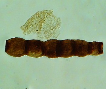
Reduviasporonites chalastus (Foster) Elsik 1999
Chain of cells.
Mean length of cells 100µm. Length of chain 330µm.
Greenland
H44/4
Reduviasporonites chalastus (Foster) Elsik 1999
Cell relatively light in colour due to the comprehensive collapse of the inner body. The very dark amorphous to granulate cell material is seen to be enveloped in the very thin, delicate inner body which is twisted and attached to the cell wall at the right terminal rim.
Length of cell 55µm.
UK
H47/2
Reduviasporonites chalastus (Foster) Elsik 1999
Three cells in a chain. Each cell with a very thin delicate outer cell wall and an inner body in various stages of shrinkage. Cell material appears to be present within the inner body.
Mean length of cells 100µm. Length of chain 330µm.
Greenland
J21/2
Reduviasporonites chalastus (Foster) Elsik 1999
Detail of specimen shown in image MFP12114_3_750_6_J21_2. Detail of cells on the left side.
Mean length of cells 100µm. Length of chain 330µm.
Greenland
J21/2
Reduviasporonites chalastus (Foster) Elsik 1999
Cell with thin outer wall; inner body absent.
Length of cell 115µm
Greenland
J36/1
Reduviasporonites chalastus (Foster) Elsik 1999
Cell lacking inner body
Cell length 63µm
UK
J39
Reduviasporonites chalastus (Foster) Elsik 1999
Well developed terminal rims and cell material. Inner body believed to be adpressed and hence optically not distinct from the main cell wall.
Length of cell 84µm
Australia
J46/2
Reduviasporonites chalastus (Foster) Elsik 1999
Chain of three short ovoid cells. Cell to the left has three terminal rims.
Mean length of cells 35µm
Saudi Arabia
K35/1
Reduviasporonites chalastus (Foster) Elsik 1999
Detail of specimen shown in image DILM_1_7827.9_K35_1. Chain of three short ovoid cells. Detail showing inner and outer cell walls. Outer cell wall approx. 1µm thick; surface smooth. Inner wall between 1 and 0.75µm thick; surface also smooth. Two walls closely adpressed.
Mean length of cells 35µm
Saudi Arabia
K35/1
Reduviasporonites chalastus (Foster) Elsik 1999
Cell wall irregularly pitted.
Length of cell 87µm.
Saudi Arabia
K40/2
Reduviasporonites chalastus (Foster) Elsik 1999
Ovoid cell with indistinct terminal rims
Length of cell 53µm
Australia
K46/1
Reduviasporonites chalastus (Foster) Elsik 1999
Cell with constricted terminal rims
Length of cell 83µm.
Saudi Arabia
K49/2
Reduviasporonites chalastus (Foster) Elsik 1999
Damaged, poor specimen
Length of cell 150µm approx.
Greenland
K51/3
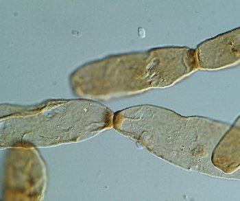
Reduviasporonites chalastus (Foster) Elsik 1999
Cells with apparent oblique connection
Cell lengths 33 and 32µm
UK
L30
Reduviasporonites chalastus (Foster) Elsik 1999
Pair of joined cells, indicating nature of cell contact and advanced state of shrinkage of the inner body in both cells.
Length of pictured cells 110 and 100µm
China
L35
Reduviasporonites chalastus (Foster) Elsik 1999
Ovoid cell with indistinct terminal rims
Length of cell 50µm
Australia
L36/4
Reduviasporonites chalastus (Foster) Elsik 1999
Flask shaped cell containing granular cell material.
Length of cell 128µm.
Russia
L49/2
Reduviasporonites chalastus (Foster) Elsik 1999
Detail of outer cell wall. Shows scabrous nature of the Moura specimens. Some evidence of pyrite deformation of the cell wall material is present.
Width of cell 47µm
Australia
M42
Reduviasporonites chalastus (Foster) Elsik 1999
Damaged, poor specimen
Length of cell 150µm approx.
Greenland
M8
Reduviasporonites chalastus (Foster) Elsik 1999
The two prominent dark cells pictured here have inner bodies which almost fill the cell cavity. The differing transparency of the cells illustrated may be due to natural variation in the thickness of one or both of the cell walls.
Cells with length between 48 and 28 µm.
Austria
N11/4
Reduviasporonites chalastus (Foster) Elsik 1999
Chain of cells with masses of cell material. The cells all have intact inner bodies that almost fill the cell cavity.
Cells with maxis lengths of 45, 28 and 32µm
Austria
N12/3
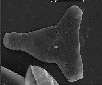
Reduviasporonites chalastus (Foster) Elsik 1999
Showing a ‘three way cell’; each terminus with a terminal rim.
Approx. length of cell 100µm
China
N21/4
Reduviasporonites chalastus (Foster) Elsik 1999
Two flask-shaped cells
Cell lengths 61 and 70µm
UK
N26/2
Reduviasporonites chalastus (Foster) Elsik 1999
Short spindle shaped cell
Length of cell 44µm
Saudi Arabia
N28
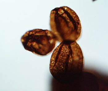
Reduviasporonites chalastus (Foster) Elsik 1999
Chain of small ovoid cells similar in morphology to the Flowerpot Formation specimens
Mean length of cells 25µm
Saudi Arabia
N30
Reduviasporonites chalastus (Foster) Elsik 1999
Ovoid cell with indistinct ovoid inner body.
Length of cell 90µm
Australia
N31
Reduviasporonites chalastus (Foster) Elsik 1999
Showing the dispersion of the cell material within the cell cavity.
Length of cell 150µm
China
N31
Reduviasporonites chalastus (Foster) Elsik 1999
Shows some of the range of cell size, shape and wall thickness within a single field of view. To the right are cells similar to those of the Greenland and Moura material. To the left are short ovoid cells.
Elongate cells length 55 and 45µm; ovoid cells have length between 29 and 45µm.
Austria
N31/3
Reduviasporonites chalastus (Foster) Elsik 1999
The cell displays a slightly shrunken inner body where the inner wall is no longer adpressed to the inner surface of the outer cell wall but has retreated a few microns. Note that the inner cell wall is still adpressed to the outer cell wall at the termini.
Main cell has length 42µm
Austria
N33/4
Reduviasporonites chalastus (Foster) Elsik 1999
Nearly rectangular outline, short cell
Length of cell 53µm
Saudi Arabia
N37/4
Reduviasporonites chalastus (Foster) Elsik 1999
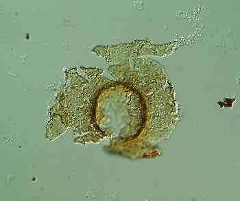
Showing a detached terminal rim. Plane of terminal face parallel to plane of slide.
Diameter of the terminal rim 27 µm
China
N40/2
Reduviasporonites chalastus (Foster) Elsik 1999
Nearly rectangular outline, short cell
Length of cell 67µm
Saudi Arabia
N41/3
Reduviasporonites chalastus (Foster) Elsik 1999
Short cell with rectangular outline and small masses of cell material
Length of cell 45µm
Saudi Arabia
N45/3
Reduviasporonites chalastus (Foster) Elsik 1999
Ovoid cell with indistinct ovoid inner body; the fold perpendicular to the long axis of the cell indicates one terminus of the inner body.
Length of cell 110µm
Australia
N46/1
Reduviasporonites chalastus (Foster) Elsik 1999
Slightly ovoid cell with indistinct inner body, though folds delineating its outline may be present. Terminal face turned towards the viewer.
Length of cell 118µm
Australia
N48/2
Reduviasporonites chalastus (Foster) Elsik 1999
Two adjacent cells; one with an inner body, one without.
Two main cells pictured have lengths of 45 and 43µm
Austria
N6
Reduviasporonites chalastus (Foster) Elsik 1999
The middle cell displays a slightly shrunken inner body where the inner wall is no longer adpressed to the inner surface of the outer cell wall but has retreated a few microns. Note that the inner cell wall is still adpressed to the outer cell wall at the termini. The lack of a strong transparency difference between parts of the cell showing the inner and outer walls indicates that the inner body has a thin wall.
Two main cells pictured have max. axis lengths of 40 and 43µm
Austria
N8
Reduviasporonites chalastus (Foster) Elsik 1999
Elongate cell with inner body in advanced state of shrinkage
Main cell length 47µm
Austria
O16/3
Reduviasporonites chalastus (Foster) Elsik 1999
Cell with constricted terminal rims
Length of cell 70µm
Saudi Arabia
O42/2
Reduviasporonites chalastus (Foster) Elsik 1999
Poorly developed ‘Y’ shaped cell
Length of cell 61µm
Saudi Arabia
O42/2
Reduviasporonites chalastus (Foster) Elsik 1999
Spindle shaped cell
Length of cell 49µm
Saudi Arabia
O44
Reduviasporonites chalastus (Foster) Elsik 1999
Very small rounded cell
Length of cell 32µm
Saudi Arabia
O45
Reduviasporonites chalastus (Foster) Elsik 1999
Cell with rectangular outline
Length of cell 69µm
Saudi Arabia
O45/2
Reduviasporonites chalastus (Foster) Elsik 1999
Short cell with rectangular outline
Length of cell 45µm
Saudi Arabia
O45/2
Reduviasporonites chalastus (Foster) Elsik 1999
Very poorly developed ‘Y’ shaped cell
Length of cell 66µm
Saudi Arabia
O47
Reduviasporonites chalastus (Foster) Elsik 1999
2 joined cells
Length of cells 44 and 67µm
Saudi Arabia
O48/1
Reduviasporonites chalastus (Foster) Elsik 1999
Flask shaped cell
Length of cell 59µm
Saudi Arabia
O48/1
Reduviasporonites chalastus (Foster) Elsik 1999
Very poorly developed ‘Y’ shaped cell
Length of cell 61µm
Saudi Arabia
O48/1
Reduviasporonites chalastus (Foster) Elsik 1999
Ovoid cell with indistinct ovoid inner body; the folds perpendicular to the long axis of the cell indicate the termini of the inner body.
Length of cell 129µm
Australia
O48/1
Reduviasporonites chalastus (Foster) Elsik 1999
Elongate cell
Length of cell 87µm
Saudi Arabia
O48/3
Reduviasporonites chalastus (Foster) Elsik 1999
Small ovoid cell with poorly developed terminal rims
Length of cell 40µm
Saudi Arabia
O49
Reduviasporonites chalastus (Foster) Elsik 1999
Small cell with rectangular outline
Length of cell 59µm
Saudi Arabia
O49
Reduviasporonites chalastus (Foster) Elsik 1999
Slightly ovoid to rectangular cell with no distinct inner body or folds; also with discontinuous terminal rims.
Length of cell 118µm
Australia
O50
Reduviasporonites chalastus (Foster) Elsik 1999
Chain of very short ovoid cells
Mean length of cells 25µm
Saudi Arabia
O51/2
Reduviasporonites chalastus (Foster) Elsik 1999
Flask shaped cell with distinct inner and outer wall layers
Length of cell 65µm
Saudi Arabia
O54/1
Reduviasporonites chalastus (Foster) Elsik 1999
Shows folds in the inner bodies of two adjacent elongate cells
Length of largest cell pictured is 65µm
Austria
P14/4
Reduviasporonites chalastus (Foster) Elsik 1999
Main cell pictured shows how shrinkage of the inner body occurs mainly in the middle parts of the cell. The inner body rarely detaches from the inner surfaces of the terminal rims unless shrinkage is very advanced.
Main cell length 43µm
Austria
P15/3
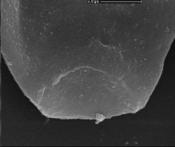
Reduviasporonites chalastus (Foster) Elsik 1999
End on view orientated normal to the terminal plane. Rim appears rectilinear in form, thickening may be incipient or as a result of folding. No pore or aperture visible in the terminal face. Terminal face planar.
Width of terminal face 20µm
Australia
P33/4
Reduviasporonites chalastus (Foster) Elsik 1999
Slightly ovoid cell with partially shrunken inner body.
Length of cell 115µm
Australia
P36
Reduviasporonites chalastus (Foster) Elsik 1999
Chain of three cells, each showing shrunken inner body with varying levels of attachment to the termini of the outer wall.
Length of pictured cells 115, 100 and 90 µm
China
P5/1
Reduviasporonites chalastus (Foster) Elsik 1999
Detail of specimen shown in image 6403380_NLD36_P5_1. Chain of three cells. Right side of chain. Illustrates attachment of the inner body to the termini of the outer wall. A helical twist may be present in the inner body of the right cell. Possibly such a twist is responsible for common diagonal folds.
Length of pictured cells 115, 100 and 90 µm
China
P5/1
Reduviasporonites chalastus (Foster) Elsik 1999
Detail of specimen shown in image 6403380_NLD36_P5_1. Chain of three cells. Left side of chain. Illustrates tenuous attachment of the inner body to the termini of the outer wall.
Length of pictured cells 115, 100 and 90 µm
China
P5/1
Reduviasporonites chalastus (Foster) Elsik 1999
Slightly ovoid cell with inner body delineated by folds.
Length of cell 92µm
Australia
P52/2
Reduviasporonites chalastus (Foster) Elsik 1999
Flask shaped cell
Length of cell 79µm
Saudi Arabia
Q30/2
Reduviasporonites chalastus (Foster) Elsik 1999
Slightly ovoid cell with distinct diagonal fold. Cf. diagonal furrow of Foster (1979).
Length of cell 100µm
Australia
R48/2
Reduviasporonites chalastus (Foster) Elsik 1999
Ovoid cell with partially shrunken inner body.
Length of cell 120µm
Australia
S36
Reduviasporonites chalastus (Foster) Elsik 1999
Cell relatively light in colour due to the comprehensive collapse of the inner body. The very dark amorphous to granulate cell material is seen to be enveloped in the very thin, delicate inner body which is twisted and attached to the cell wall at both ends. At the right is a strand of material which may represent the shrunken inner body of a previously adjacent cell.
Length of cell 50µm.
UK
S39/1
Reduviasporonites chalastus (Foster) Elsik 1999
Rectangular, elongate cell with indistinct inner body.
Length of cell 130µm
Australia
S44
Reduviasporonites chalastus (Foster) Elsik 1999
Discontinuous terminal rim present; inner body not apparent.
Length of cell 62µm
Australia
Single grain mount
Reduviasporonites chalastus (Foster) Elsik 1999
Discontinuous terminal rim present; inner body not apparent. Left side shows incipient weakness of the outer wall shown by separation of the rim from the rest of the wall. Cell material distinct, granular in appearance.
Length of cell 56µm
Australia
Single grain mount
Reduviasporonites chalastus (Foster) Elsik 1999
Detail of specimen shown in image BILLA_262m_SGM_3_2. Discontinuous terminal rim present; incipient weakness of the outer wall shown by separation of the rim from the rest of the wall
Length of cell 83µm
Australia
Single grain mount
Reduviasporonites chalastus (Foster) Elsik 1999
Discontinuous terminal rim present; incipient weakness of the outer wall shown by separation of the rim from the rest of the wall
Length of cell 83µm
Australia
Single grain mount
Reduviasporonites chalastus (Foster) Elsik 1999
Discontinuous terminal rim present; incipient weakness of the outer wall shown by separation of the rim from the rest of the wall
Length of cell 57µm
Australia
Single grain mount
Reduviasporonites chalastus (Foster) Elsik 1999
Discontinuous terminal rim present
Length of cell 99µm
Australia
Single grain mount
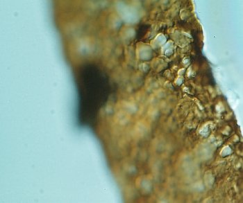
Reduviasporonites chalastus (Foster) Elsik 1999
Detail of specimen shown in image BILLA_262m_SGM_4. Showing granular cell material.
Length of cell 99µm
Australia
Single grain mount
Reduviasporonites chalastus (Foster) Elsik 1999
To the right of the picture the inner body is visible, closely adpressed to the inner surface of the cell wall. Granular cell material present.
Length of cell 102µm
Australia
Single grain mount
Reduviasporonites chalastus (Foster) Elsik 1999
Detail of specimen shown in image BILLA_262m_SGM_5. Inner body can be seen folded back approximately half way along the cell. This may account for lighter colour in the left side of the cell.
Length of cell 102µm
Australia
Single grain mount
Reduviasporonites chalastus (Foster) Elsik 1999
Detail of specimen showing a complete cell joined to a partial cell that has broken at the weakness at the base of the terminal rim. Illustrates the fine suture at the join between the adjacent terminal rims.
See scale bar
Austria
Stub 1
Reduviasporonites chalastus (Foster) Elsik 1999
Chain of three cells with deeply concave compressional shape.
See scale bar
Austria
Stub 1
Reduviasporonites chalastus (Foster) Elsik 1999
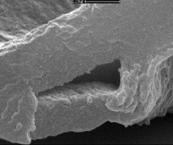
Detail of specimen showing a complete cell joined to a partial cell. Longitudinal tears in the outer cell wall are present.
See scale bar
Austria
Stub 1
Reduviasporonites chalastus (Foster) Elsik 1999
Detail of specimen above. Wall of inner body may be visible within the longitudinal tear.
See scale bar
Austria
Stub 1
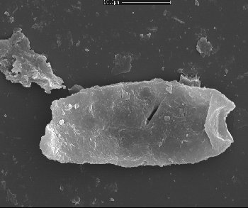
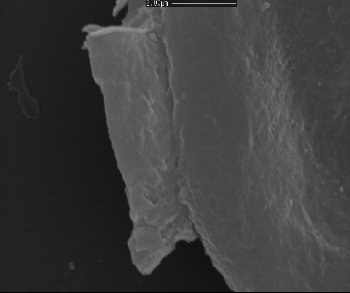
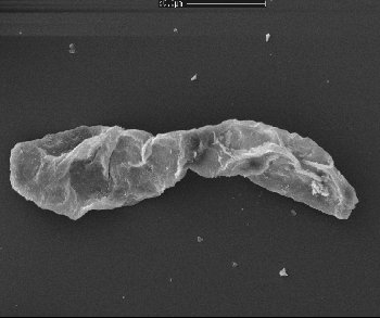
Reduviasporonites chalastus (Foster) Elsik 1999
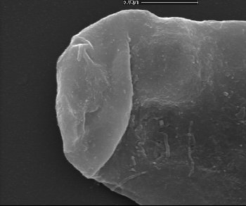
Elongate cell with well developed terminal rim.
See scale bar
Austria
Stub 1
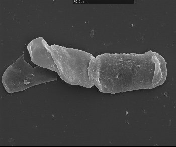
Reduviasporonites chalastus (Foster) Elsik 1999
Chain of two cells. Left cell has a diagonal fold due to twisting. Illustrates the rigidity of the terminal rims in comparison with the flaccidity of the otherwise unsupported cell wall.
See scale bar
Austria
Stub 1
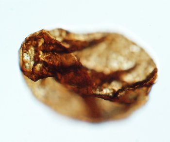
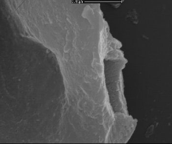
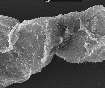
Reduviasporonites chalastus (Foster) Elsik 1999
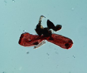
This specimen illustrates the characteristic ‘Christmas cracker’ morphology of cell chains after vigorous ultrasound treatment. The central cell shown is intact but the two adjacent cells are broken at the base of their respective terminal rims. This illustrates the comparative strength of the bonds between terminal rims.
See scale bar
Austria
Stub 1
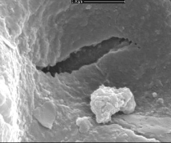
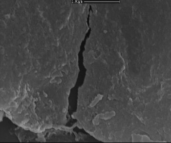
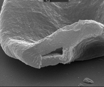
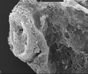
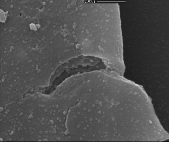
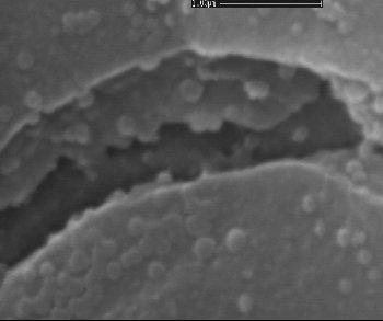
Reduviasporonites chalastus (Foster) Elsik 1999
Crack in cell wall. Illustrates the lack of a clear inner body cell wall and, possibly, that in this specimen, the inner body is tightly adpressed to the main cell wall.
See scale bar
Austria
Stub 1
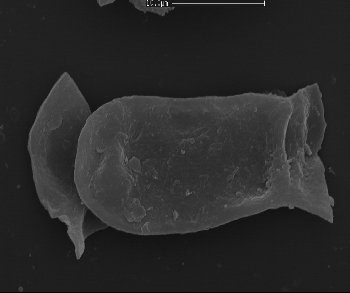
Reduviasporonites chalastus (Foster) Elsik 1999
Chain of two cells. Left cell has ‘Christmas cracker’ morphology.
See scale bar
Austria
Stub 1
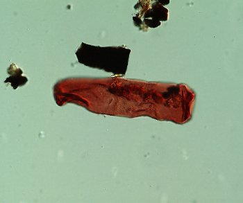
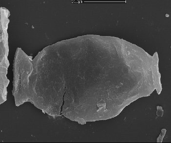
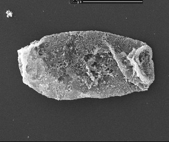
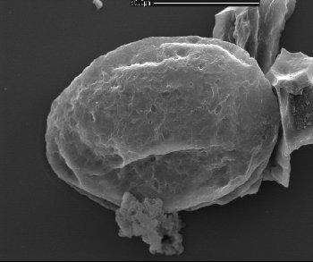
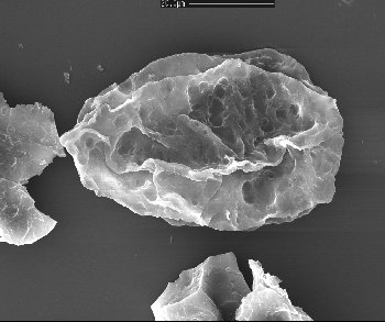
Reduviasporonites chalastus (Foster) Elsik 1999
Large, elongate cell.
See scale bar
Austria
Stub 1
Reduviasporonites chalastus (Foster) Elsik 1999
Chain of two cells, left cell broken. In overall size (right cell less than 20µm in length) and morphology, these cells are similar to those described by Wilson (1962) and assigned to R. catenulatus.
See scale bar
Austria
Stub 1
Reduviasporonites chalastus (Foster) Elsik 1999
This specimen illustrates the characteristic ‘Christmas cracker’ morphology of cell chains after vigorous ultrasound treatment. The central cell shown is intact but the two adjacent cells are broken at the base of their respective terminal rims. This illustrates the comparative strength of the bonds between terminal rims.
See scale bar
Austria
Stub 1
Reduviasporonites chalastus (Foster) Elsik 1999
One intact cell with adjacent partial cells. Right side shows detachment of two terminal rims.
See scale bar
Austria
Stub 1
Reduviasporonites chalastus (Foster) Elsik 1999
Detail of detachment of two terminal rims.
See scale bar
Austria
Stub 1
Reduviasporonites chalastus (Foster) Elsik 1999
Cell with an oval outline, probably originally ovoid in shape with adjacent partial cell. Right side shows a break in the cell wall.
See scale bar
Austria
Stub 1
Reduviasporonites chalastus (Foster) Elsik 1999
Detail of the above specimen (right side) showing the inner surface of ?one of the cell walls.
See scale bar
Austria
Stub 1
Reduviasporonites chalastus (Foster) Elsik 1999
Detail of the above specimen (left side).
See scale bar
Austria
Stub 1
Reduviasporonites chalastus (Foster) Elsik 1999
This specimen illustrates the characteristic ‘Christmas cracker’ morphology. On the right the two tightly connected terminal rims can be seen.
See scale bar
Austria
Stub 1
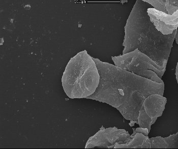
Reduviasporonites chalastus (Foster) Elsik 1999
Elongate cell with terminal rim turned toward the viewer.
See scale bar
Austria
Stub 1
Reduviasporonites chalastus (Foster) Elsik 1999
Detail of the above specimen (left side) with terminal rim turned toward the viewer.
See scale bar
Austria
Stub 1
Reduviasporonites chalastus (Foster) Elsik 1999
Two ?recently detached cells. Right cell shows possible development of a third terminal rim.
See scale bar
Austria
Stub 1
Reduviasporonites chalastus (Foster) Elsik 1999
Detail of the above specimen (upper part) with terminal rim.
See scale bar
Austria
Stub 1
Reduviasporonites chalastus (Foster) Elsik 1999
Detail of terminal rim. Central smooth area may correspond to central ‘boss’ of image ‘DILM_1_7827.9_Stub6_2.jpg’ from Saudi Arabia sample DILM-1 7827.
See scale bar
Austria
Stub 1
Reduviasporonites chalastus (Foster) Elsik 1999
Chain of two tightly connected cells. Left cell in addition shows connection to the remains of an adjacent cell.
See scale bar
Austria
Stub 1
Reduviasporonites chalastus (Foster) Elsik 1999
Detail of the above chain (left side).
See scale bar
Austria
Stub 1
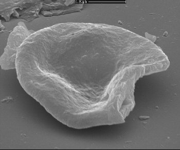
Reduviasporonites chalastus (Foster) Elsik 1999
Oblique image of specimen shown in image ‘6403434_2_T1_Stub1_15.tif’ (Austria T1 6403434 2 Stub 1). Shows 3D shape of the compressed cell. The extent and pattern of folding indicates that the cell was originally ovoid in shape.
See scale bar
Austria
Stub 1
Reduviasporonites chalastus (Foster) Elsik 1999
Oblique image of specimen shown in image ‘6403434_2_T1_Stub1_15.tif’ (Austria T1 6403434 2 Stub 1). Detail.
See scale bar
Austria
Stub 1
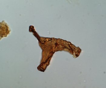
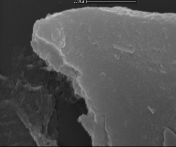
Reduviasporonites chalastus (Foster) Elsik 1999
Oblique image of specimen shown in image ‘6403434_2_T1_Stub1_15.tif’ (Austria T1 6403434 2 Stub 1). Detail.
See scale bar
Austria
Stub 1
Reduviasporonites chalastus (Foster) Elsik 1999
Oblique image of specimen shown in image ‘6403434_2_T1_Stub1_15.tif’ (Austria T1 6403434 2 Stub 1). Detail illustrates the thickness (approx. 0.75µm), and apparently homogeneous nature of the cell wall. Area within presumably represents the internal cavity of the cell. The inner surface of the wall illustrated appears to be striated.
See scale bar
Austria
Stub 1
Reduviasporonites chalastus (Foster) Elsik 1999
Oblique image of specimens.
See scale bar
Austria
Stub 1
Reduviasporonites chalastus (Foster) Elsik 1999
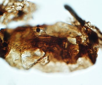
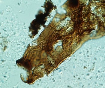
Poorly preserved elongate cell. Terminal rim to the left has poorly developed raised circular boss approximately 3 µm wide
See scale bar
Austria
Stub 12
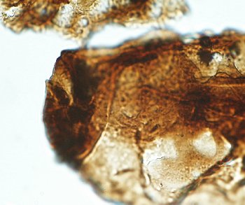
Reduviasporonites chalastus (Foster) Elsik 1999
Poorly preserved elongate cell. Terminal rim detail.
See scale bar
Austria
Stub 12
Reduviasporonites chalastus (Foster) Elsik 1999
Chain of 6 cells. Several of the cells have partially collapsed walls between the terminal rims suggesting that the latter are the most rigid part of the cell structure.
See scale bar
Austria
Stub 12
Reduviasporonites chalastus (Foster) Elsik 1999
Detail of terminal rim
See scale bar
Austria
Stub 12
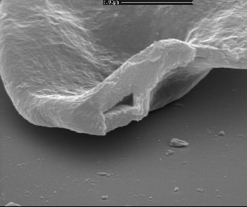
Reduviasporonites chalastus (Foster) Elsik 1999
Three way cell and ?recently detached elongate cell. Left terminal rim has poorly developed raised circular boss approximately 2 µm wide
See scale bar
Austria
Stub 12
Reduviasporonites chalastus (Foster) Elsik 1999
Detail of terminal rim of elongate cell in the above.
See scale bar
Austria
Stub 12
Reduviasporonites chalastus (Foster) Elsik 1999
Flask shaped cell with well developed left terminal rim. Specimen has corroded outer cell wall.
See scale bar
China
Stub 13
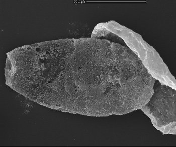
Reduviasporonites chalastus (Foster) Elsik 1999
Pair of joined cells.
See scale bar
China
Stub 13
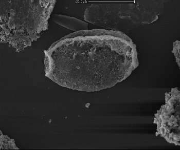
Reduviasporonites chalastus (Foster) Elsik 1999
Pair of joined cells. Detail of the join between the cells. Join is marked by a fine suture.
See scale bar
China
Stub 13
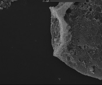
Reduviasporonites chalastus (Foster) Elsik 1999
Large rounded cell with well developed terminal rims. Specimen has corroded outer cell wall.
See scale bar
China
Stub 13
Reduviasporonites chalastus (Foster) Elsik 1999
Rounded cell with well developed terminal rims. Specimen has corroded outer cell wall and prominent longitudinal fold.
See scale bar
China
Stub 13
Reduviasporonites chalastus (Foster) Elsik 1999
Rounded cell with well developed terminal rims. Detail of left terminal rim.
See scale bar
China
Stub 13
Reduviasporonites chalastus (Foster) Elsik 1999
Rounded cell with well developed terminal rims.
See scale bar
China
Stub 13
Reduviasporonites chalastus (Foster) Elsik 1999
Detail of terminal rim. Fragment of previously attached cell still present.
See scale bar
China
Stub 13
Reduviasporonites chalastus (Foster) Elsik 1999
Elongate cell with well developed diagonal fold.
See scale bar
China
Stub 13
Reduviasporonites chalastus (Foster) Elsik 1999
Elongate cell with well developed diagonal fold. The right terminal rim is partially detached along the incipient weakness in the cell wall at the base of the terminal rim. The left terminal rim is very well developed.
See scale bar
China
Stub 13
Reduviasporonites chalastus (Foster) Elsik 1999
Detail of left terminal rim. Area enclosed has a hole in the outer cell wall, probably too regular to be a pore or aperture.
See scale bar
China
Stub 13
Reduviasporonites chalastus (Foster) Elsik 1999
Cell with well developed longitudinal fold. This and other specimens indicate a cell wall corrosion form that is similar in appearance to ‘exfoliation’. That the wall appears to ‘flake off’ in this way suggests that it may be lamellate in structure.
See scale bar
China
Stub 13
Reduviasporonites chalastus (Foster) Elsik 1999
Small rounded cell with diagonal fold possibly caused by twisting movement.
See scale bar
Saudi Arabia
Stub 6
Reduviasporonites chalastus (Foster) Elsik 1999
Detail of terminal rim showing raised rim area and enclosed central, circular depressed area. The central part of the depressed area has a raised circular boss approximately 2 µm wide.
See scale bar
Saudi Arabia
Stub 6
Reduviasporonites chalastus (Foster) Elsik 1999
Three way cell. Upper terminal rim is slightly folded over.
See scale bar
Saudi Arabia
Stub 6
Reduviasporonites chalastus (Foster) Elsik 1999
Right side illustrates tear in the wall just at the base of the terminal rim. Suggests as do other light microscope images, that there is an incipient weakness at the base of the terminal rim.
See scale bar
Saudi Arabia
Stub 6
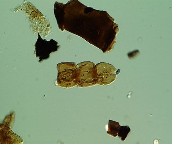
Reduviasporonites chalastus (Foster) Elsik 1999
Chain of 5 cells. Several of the cells have partially collapsed walls between the terminal rims, suggesting that the latter are the most rigid part of the cell structure. The second cell from the right is a 3 way cell.
See scale bar
Saudi Arabia
Stub 6
Reduviasporonites chalastus (Foster) Elsik 1999
Chain of 5 cells above. Detail of terminal rim.
See scale bar
Saudi Arabia
Stub 6
Reduviasporonites chalastus (Foster) Elsik 1999
Spindle shaped cell with tear in the outer wall.
See scale bar
Saudi Arabia
Stub 6
Reduviasporonites chalastus (Foster) Elsik 1999
Detail of outer cell wall tear. Shows the outer wall thickness (approx. 0.5µm) and the outer surface of the inner wall beneath.
See scale bar
Saudi Arabia
Stub 6
Reduviasporonites chalastus (Foster) Elsik 1999
Detailed close up of outer cell wall tear.
See scale bar
Saudi Arabia
Stub 6
Reduviasporonites chalastus (Foster) Elsik 1999
Rounded cell with poorly developed terminal rim on the left. Specimen has corroded outer cell wall.
See scale bar
Australia
Stub 8
Reduviasporonites chalastus (Foster) Elsik 1999
Rounded cell; no distinct terminal rims. Specimen has corroded outer cell wall.
See scale bar
Australia
Stub 8
Reduviasporonites chalastus (Foster) Elsik 1999
Rounded cell with poorly developed terminal rim on the left. Specimen has corroded outer cell wall.
See scale bar
Australia
Stub 8
Reduviasporonites chalastus (Foster) Elsik 1999
Long cell with semi transparent outer cell wall
Length of cell 100µm
Saudi Arabia
T37/2
Reduviasporonites chalastus (Foster) Elsik 1999
Two joined ovoid cells. Cf. B. helbyi f. gregata
Mean length of cells 45µm.
UK
T44/3
Reduviasporonites chalastus (Foster) Elsik 1999
Shrunken and twisted inner body envelopes a mass of dense granulate cell material. Mass and enveloping inner body forms a spindle shape. Termini of inner body still attached to the inner surfaces of the terminal rims.
Length of cell 75µm.
Russia
T55/1
Reduviasporonites chalastus (Foster) Elsik 1999
Ovoid cell with rectilinear folds possibly in the inner body wall. Cf. diagonal furrow of Foster (1979).
Length of cell 120µm
Australia
U42
Reduviasporonites chalastus (Foster) Elsik 1999
Group of ovoid cells with regular folds indicating approx. position and shape of the inner body. Cells joined where termini of inner body reach inner surface of the outer body.
Approx. mean length of cells 80µm
Australia
U46/3
Reduviasporonites chalastus (Foster) Elsik 1999
Cells with circular outline.
Length of cells 53 and 50µm.
UK
U51/1
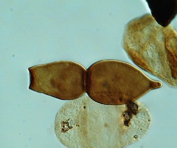
Reduviasporonites chalastus (Foster) Elsik 1999
Flask and spindle shaped cells joined
Length of cells 57 and 46µm
Saudi Arabia
W33
Reduviasporonites chalastus (Foster) Elsik 1999
Ovoid cell with rectilinear folds possibly in the inner body wall. Cf. diagonal furrow of Foster (1979).
Length of cell 120µm
Australia
Y47
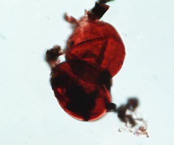
Reduviasporonites chalastus (Foster) Elsik 1999
Chain with apparent pendant inner body still attached to the adjacent cell
Mean length of cells 45µm
Saudi Arabia
Y48