Fossil Taxonomy – Carboniferous spores


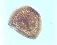
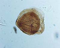
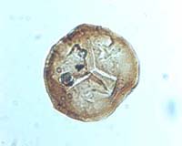
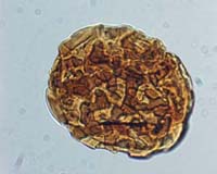
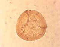
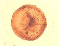
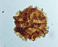
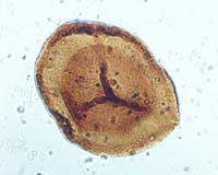
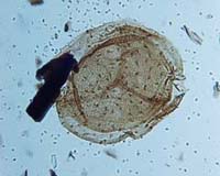
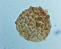
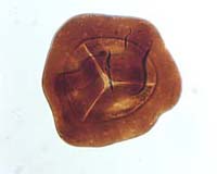
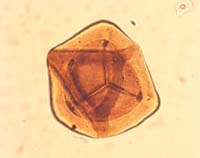
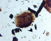
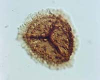
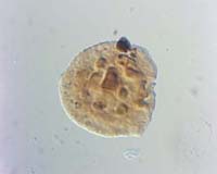
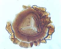
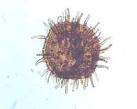
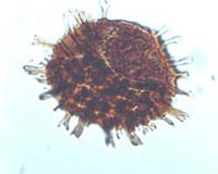
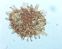
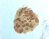
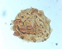
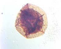
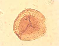
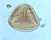
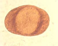
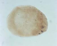
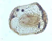
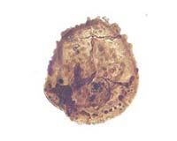
Angulisporites splendidus Bhardwaj, 1954
Radial, trilete, camerate miospores. Amb rounded triangular to subcircular or oval. Miospores consist of inner body conformable in outline with equatorial margin, surrounded by exoexinal body with thickened equatorial rim approximately 5 µm wide. Trilete mark distinct, sutures straight, extending at least as far as the margin of the inner body; simple or accompanied by relatively low elevated, flexuous folds of the exoexine that extend decreasing in height to the equatorial margin of spore. Intexine relatively thick; forms inner body which occupies between two-thirds and three-quarters of the spore diameter. Exoexine thicker; extent of separation of exoexine from intexine difficult to ascertain but inner body commonly possesses independent compression folds suggesting separation in, at least, the equatorial region and part of the distal surface. Exoexine has thickened equatorial rim, similar to a cingulum, which occupies up to half the width of exoexinal extension. Exoexine infrapunctate.
Bhardwaj, 1954, p. 516, text-fig. 4
Overall equatorial diameter 70(83)105 µm. Diameter of inner body 50(64)76 µm
The presence of the cingulum-like thickening at the equatorial margin of the exoexinal body distinguishes representatives of this genus from Endosporites.
Late Pennsylvanian to Early Permian; [latest Westphalian D (Asturian) to early Autunian]; VC Biozone (Sánchez de Posada et al., 2002; Clayton et al., 1977)

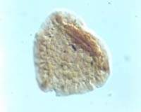
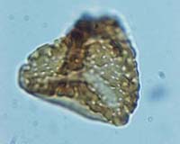
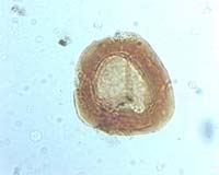
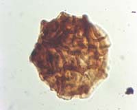
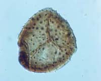
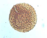
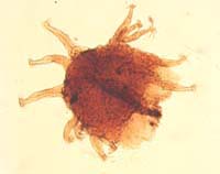
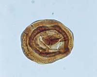
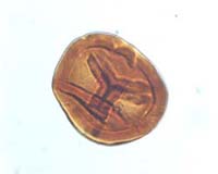
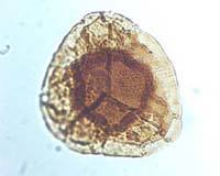
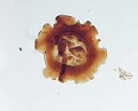
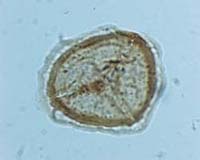
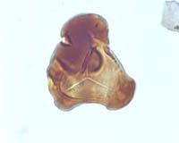
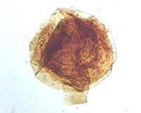
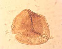
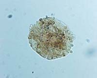
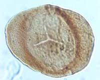
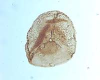
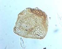
Auroraspora macra Sullivan, 1968
Radial, trilete, camerate miospores. Amb circular to subcircular, frequently irregular due to folding. Trilete mark distinct, sutures straight, extend at least two-thirds of the radius of the inner body, commonly accompanied by low thickened labra with irregular outer margins, which extend to the equatorial margin of spore. Intexine thin forming well defined inner body. Exoexine thin, variably separated from intexine, commonly folded, densely infrapunctate. Exoexine thickened in the equatorial plane to form limbus-like structure of variable width.
Sullivan, 1968, pp. 124–125, pl. 27, figs. 6–10. Clayton, 1971, p. 580, pl. 1, fig. 3. Higgs et al., 1988, p. 69, pl. 9, figs 17–19
Overall equatorial diameter 48(58)78 µm. Diameter of inner body 26(31)35 µm
The distinctive thickened equatorial margin of the exoexine renders this species readily distinguishable from all other taxa assigned to the genus. Auroraspora solisortus Hoffmeister, Staplin & Malloy 1955 is larger, lacks the thickened exoexinal equatorial rim and possesses a radially oriented series of exoexinal folds. A. hyalina (Naumova) Streel in Becker et al. 1974 is slightly smaller in size and possesses a more filmy exoexine.
Uppermost Devonian to Mississippian; Strunian to early Visean; LL to Pu biozones (Higgs et al., 1988)
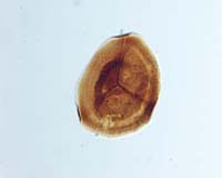
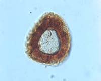
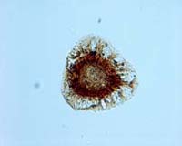
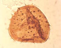
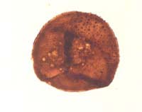
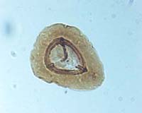
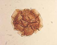
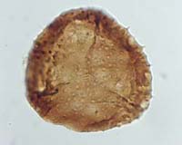
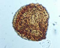
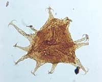
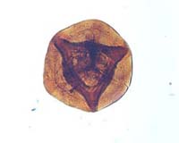
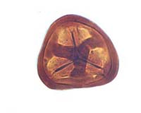
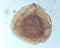
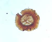
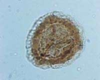
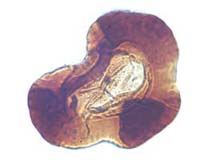
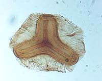
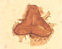
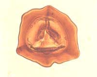
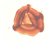
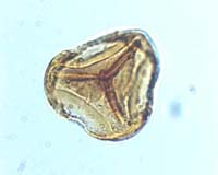
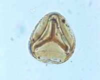
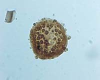
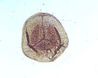
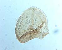
Bellispores nitidus (Horst) Sullivan, 1964
Radial, trilete, acamerate, cingulate miospores. Amb triangular with concave or more rarely straight sides and rounded apices. Outline crenulate. Trilete mark distinct, sutures straight, simple or with low narrow thickened labra, extending to the inner margin of the cingulum. Intexine thin, forming wall of spore body. Exoexine closely adpressed over both proximal and distal surfaces but extended in the equatorial plane to form irregular narrow cingulum, 2–5 µm wide, with irregular crenulate margin but of more or less uniform thickness. Distal surface ornamented with thickened Y-shaped structure with the three components extending from the distal pole to the cingulum which they join in the radial positions. Margins of distal thickened ribs crenulate in the same style as the cingulum, individual ribs up to 10 µm wide. Proximal surface laevigate, distal surface including distal thickened ribs and the cingulum foveolate, with foveolae less than 2 µm in diameter. Foveolae may be randomly distributed or arranged in rows. Distal thickened ribs often bear two rows of foveolae running down their length.
Horst, 1955, p. 181, p1. 24, fig. 81. Artüz, 1957, p. 255, pl. 7, fig. 49. Sullivan, 1964, p. 375
Overall equatorial diameter 27(36)45 µm
Smith and Butterworth (1967) suggested that Bellispores bellis Artüz 1957 and Simozonotriletes trilinearis Artüz 1957 are synonymous with Bellispores nitidus.
Mississippian to Pennsylvanian; late Visean [Brigantian] to Bashkirian [early Duckmantian]; VF to NJ biozones (Clayton et al., 1977; Owens et al. 2004)
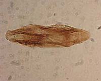
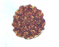
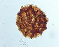
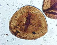
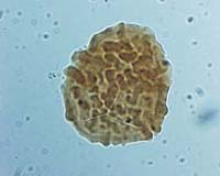
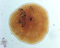
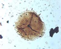
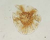
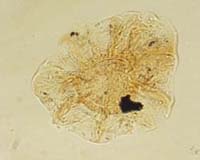
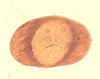
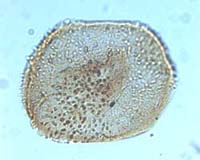
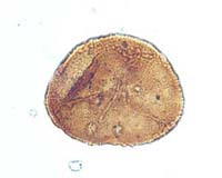
Cheiledonites major Doubinger, 1957
Pollen grains with oval or fusiform outline. Specimens characterised by well-developed furrow which extends almost the complete length of the grain. Furrow sometimes contorted by folding, or accompanied on either side by thickened lip-like folds. Remainder of exine also folded with axes of irregular folds parallel to long axis of grain. Exine surface finely infrapunctate.
Doubinger, 1957, pp. 125–133, fig. 3
Length of grain 35–42 µm Width of grain 18–20 µm.
Few formal descriptions of this species exist. The specimens described by Doubinger (1957) were recovered from Stephanian B sediments from Decazeville, France. The material documented in the present study is from Stephanian C–D sediments in north-west France, and appears to be closely comparable to Doubinger’s material in all but one respect, size. The present specimens have a maximum length of 120 µm.
The longitudinal furrow bordered by narrow folds distinguishes this species from representatives of the genus Entylissa. Cheiledonites potoniei Doubinger 1957, also described from Stephanian B sediments from Decazeville, France is distinguished by its smaller size.
Late Pennsylvanian to Permian; late Kazimovian [late Stephanian A (Barruelian)] to ?Early Permian; ST Biozone (Clayton et al., 1977)
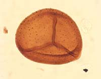
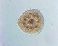
Cingulizonates bialatus (Waltz) Smith & Butterworth, 1967
Radial, trilete, cingulate miospores. Amb rounded triangular to subcircular. Trilete mark usually obscure, occasionally distinct, sutures simple, straight, extending to margin of inner body. Intexine thin, laevigate, forming rounded triangular or subcircular inner body, Exoexine extending in the equatorial plane to form bizonate cingulum. Inner zone thicker and wider than outer zone; inner zone overlaps the equatorial margin of the inner body on the distal surface. Outer margin of inner thicker zone bears numerous tubercular processes that project across outer thinner zone. Numerous radially oriented thickened ribs or plications extend across both zones of cingulum. Thinner outer zone of cingulum may be ornamented on its distal surface with small coni less than 1 µm in height.
Smith and Butterworth, 1967, pp. 260–261, pl. 21, figs. 3–4
Overall equatorial diameter 25(54)77 µm. Diameter of inner body 20(27)35 µm
The specimens originally described by Butterworth and Williams as Densosporites striatus were transferred to C. bialatus by Smith and Butterworth (1967). Cingulizonates loricatus (Loose) Butterworth & Smith 1964 is similar but lacks the development of tubercles on the outer margin of the inner thick zone of the cingulum.
Mississippian to Pennsylvanian; Visean to Bashkirian [Duckmantian]; VF to NJ biozones (unpublished information)
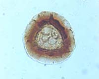
Cingulizonates capistratus (Hoffmeister, Staplin & Malloy) Staplin & Jansonius, 1974
Radial, trilete, cingulate miospores. Amb convexly rounded triangular with rounded or roundly pointed apices. Trilete mark distinct to obscure, sutures straight, simple or accompanied by low, narrow. thickened labra, extending to equatorial margin of inner body. Intexine thin, laevigate, forming rounded triangular inner body. Exoexine normally closely adpressed to intexine and extended in the equatorial plane to form differentially thickened cingulum. Minor separation may exist in the equatorial plane between the intexine and exoexine. Cingulum bizonate, consisting of narrow, thick, inner zone and wider, thin, outer zone. Thin outer zone divisible into two components: an inner section containing a large number of radially orientated elongate vacuoles separated by narrow walls, and an outer part that is weakly striated and may bear an ornament of small coni. Cingulum approximately half spore radius in width.
Smith and Butterworth, 1967, pp. 261–262, pl. 21, figs. 5–6
Overall equatorial diameter 41(51)60 µm
Radiizonates aligerens (Knox) Staplin & Jansonius 1964 differs in having longer trilete rays and a wider cingulum which has more prominent radiating struts separating internal vacuoles.
Brindley and Spinner (1989) noted the onset of common Cingulizonates cf. capistratus sensu Smith and Butterworth 1967 at a level within the NC Biozone, which they considered to coincide with the Visean–Namurian boundary in sequences of the Midland Valley of Scotland, northern England and Northern Ireland. However, Owens et al. (2004) have shown that the onset of common C. cf. capistratus occurs within the late Visean [Brigantian].
(Cingulizonates cf. capistratus sensu Smith & Butterworth 1967) Mississippian; late Visean to early Serpukhovian [early Arnsbergian]; Cc to TK biozones (Owens et al., 2004).
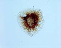
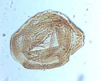
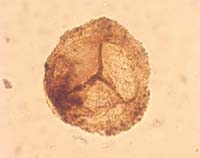
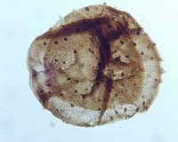
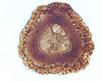
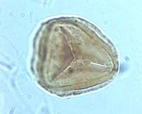
Cirratriradites saturni (Ibrahim) Schopf, Wilson & Bentall, 1944
Radial, trilete, cingulizonate, camerate miospores. Amb rounded triangular to subcircular. Trilete mark distinct, sutures straight, extending to the equatorial margin of inner body, often obscured by elevated flexuous folds of the exoexine that extend decreasing in height to the equator of spore. Intexine thin, granulate, forming rounded triangular inner body. Exoexine relatively thick, extended in the equatorial plane to form differentially thickened zona consisting of two parts, an inner thickened part which appears to overlap the margin of the inner body on the distal surface and a wider thinner outer part which frequently appears faintly radially striated. Thinner outer section of zona may contain small radially orientated internal vacuoles. Distal exoexine bears prominent foveola in the polar region. Margin of foveola thickened to produce low thickened rim. A single foveola may be present with a circular or ‘clover leaf’ shape. Specimens with up to three separate distal foveolae also occur.
Smith and Butterworth, 1967, pp. 258–259, pl. 21, figs. 1–2
Overall equatorial diameter 59(73)100 µm. Diameter of inner body 45(58)79 µm
Distinguished from other taxa assigned to the genus by the presence of a broad striate zona and one to three distal fovea. Smith and Butterworth (1967) considered that Cirratriradites flabelliformis Wilson & Kosanke was synonymous with C. saturni. C. flabelliformis was originally described as having three foveolae but Smith and Butterworth (1967) provided evidence that complete gradation exists between forms with one to three foveolae.
Late Mississippian to Pennsylvanian; Serpukhovian [Pendleian] to Kazimovian, [Stephanian A (Barruelian)]; NC to ST biozones (Clayton et al., 1977)
Colatisporites decorus (Bharadwaj & Venkatachala) Williams in Neves et al., 1973
Radial, trilete, camerate miospores. Amb circular, subcircular or rounded triangular. Trilete mark distinct or indistinct, sutures extending to margin of intexine; straight, simple or accompanied by elevated narrow folds or labra, extending to or almost to the equatorial margin where they fuse laterally with the curvaturae. Intexine sometimes clearly separated from the exoexine, otherwise closely adpressed. Intexine may form indistinct inner body occupying at least four-fifths of the spore diameter. Degree of cameration variable but exoexine probably attached to intexine on both proximal and distal surfaces. Exoexine relatively thick, strongly infrapunctate.
Bharadwaj and Venkatachala, 1961, p. 39, pl. 10, figs. 142–164. Williams in Neves et al., 1973, p. 41, pl. 2, figs. 11–13
Overall equatorial diameter 40(53)75 µm
Colatisporites denticulatus Neville in Neves et al., 1973 differs by possessing an exoexinal ornament of small spinae, bacula, coni and grana.
Mississippian; Tournaisian; PC to CM biozones (Higgs et al., 1988)
Convolutispora circumvallata Clayton, 1971
Radial trilete acamerate miospores. Amb circular–subcircular. Trilete mark distinct or obscured by ornamentation, sutures simple or accompanied by wide labra, extending more than three-quarters of spore radius. Ornament on entire distal surface and equatorial portion of proximal surface consists of rounded anastomosing ridges, some of which may be fused together to form an imperfect reticulum. Ridges variable in width along length and often have expanded tops particularly where ridges join. The channels and irregular lumina between the ridges are normally equal to or wider than the mean width of the ridges. Ridges 3–12 µm high and 5–15 µm wide. Exine up to 5 µm thick.
Clayton, 1971, p. 582, pl. 1, figs. 5–7. Higgs, Clayton and Keegan, 1988, p. 63, pl. 6, fig. 12
Overall equatorial diameter 64(90)112 µm
The range of morphological variability displayed by this species is wide but few specimens display a sufficiently well developed reticulation to justify inclusion within the genus Dictyotriletes. Reticulatisporites ancoralis Balme & Hassell 1962 is superficially similar but has a better developed reticulum and a thinner exine.
Mississippian; Tournaisian to Visean; CM to Pu biozones (Clayton et al., 1977)
Convolutispora major (Kedo) Turnau, 1978
Radial, trilete miospores. Amb rounded triangular to subcircular, equatorial margin undulate. Trilete mark distinct to indistinct; often obscured by ornamentation. Sutures straight, accompanied by low broad labra, up to 8 µm wide, extending at least 50 % of spore radius and sometimes reaching almost to equatorial margin. Exine up to 11 µm thick (including ornament). Distal surface and more equatorial portion of proximal surface ornamented by dense irregular, imperfect reticulum of anastomosing variably thickened ridges, individual ridges 6–10 µm wide, 6 µm high, rounded in profile. Lumina irregular, branched, 3–10 µm wide. Ridges on equatorial portion of proximal surface shorter and occasionally wart-like. Contact areas often smooth.
Turnau, 1978, p. 8, pl. 2, figs. 18a, b and 19
Equatorial diameter 69(95)119 µm
The coarse ornament of convolute ridges forming an imperfect reticulum distinguishes this species from others assigned to Convolutispora. Convolutispora harlandi Playford 1962 has coarse convolute ridges but these are irregularly and densely arranged and do not form a reticulum. C. labiata Playford 1962 has similar broad, low, thickened labra accompanying the trilete sutures, but differs from C. major in possessing an ornament of fine convolute ridges.
Mississippian; Tournaisian (Turnau, 1978).
Crassispora aculeata Neville, 1968
Radial, trilete miospores. Amb subcircular to rounded triangular. Trilete mark distinct, sutures straight, extending to or nearly to the spore margin; accompanied by elevated flexuous folds that extend, decreasing in height, to the inner margin of the equatorial crassitude with which they fuse laterally. Apical papillae common in the intertectal areas. Exine laevigate or infrapunctate, thickened in the equatorial region to form ill-defined crassitude with weak inner margin. Crassitude invaginated in the radial positions where it fuses with folds accompanying trilete sutures to form clearly defined curvaturae which run just on the proximal side of the equatorial margin. Distal surface of exine and equatorial margin ornamented with spinae and rare coni; elements 1.5–15 µm in height, and 1–5.5 µm in basal diameter. Approximately 20 elements project at the equatorial margin, elements normally discrete and more or less constant in size on individual specimens.
Neville, 1968, pp. 443–446, pl. 2, figs. 5–6
Overall equatorial diameter 56(74)96 µm
Crassispora maculosa (Knox) Sullivan 1964 is similar in size, but is distinguished by possessing an ornament of more densely distributed smaller coni.
Mississippian; Visean to early Serpukhovian [Pendleian]; TC to NC Biozones (Clayton et al., 1977).
Crassispora kosankei (Potonié & Kremp) Bharadwaj, 1957
Radial, trilete, camerate-acamerate miospores. Amb circular to subcircular or oval. Trilete mark distinct to indistinct; rays either simple, straight, extending almost to the inner margin of the equatorial crassitude or split open to form triangular tear in the polar region. Exine characteristically thickened in the equatorial region to form crassitude; inner margin of crassitude ill-defined and commonly invaginated in radial positions. Distal surface of exine ornamented with numerous discrete coni up to 2 µm height and basal diameter. Elements project at equatorial margin but appear absent from the proximal surface. Apical papillae commonly present. Exine surface between coni, densely infrapunctate. Secondary folding common.
Potonié and Kremp, 1955, p. 71, pl. 13, figs. 208–213. Smith and Butterworth, 1967, pp. 254–255, p1. 19, figs. 2–4
Overall equatorial diameter 40(64)105 µm
Crassispora ovalis Bharadwaj 1957 originally distinguished only by size from Crassispora kosankei, was incorporated within the circumscription of the latter species by emendation of Smith and Butterworth (1967). The size ranges recorded for different populations of the species are known to reflect different oxidation procedures.
Owens et al. (1977) and Clayton et al. (1977) considered that Crassispora kosankei becomes abundant at the base of the KV Biozone, approximately at the Alportian–Kinderscoutian boundary, being infrequent or rare in the earliest Namurian. Later Owens in Ramsbottom (1981) reported abundant C. kosankei in palynomorph assemblages from the boundary stratotype section of the Chokierian.
Late Mississippian to Pennsylvanian; Serpukhovian to Kasimovian [Pendleian to Stephanian]; NC to NBM biozones (Clayton et al., 1977)
Crassispora maculosa (Knox) Sullivan, 1964
Radial, trilete, camerate miospores. Amb circular to subcircular. Trilete mark distinct, sutures straight, extending half to three-quarters of spore radius, accompanied by low relatively narrow, thickened labra, individually up to 4 µm wide, which fuse equatorially into the variably defined equatorial crassitude which is frequently slightly invaginated in the radial positions. Crassitude narrow, weakly developed with ill-defined inner margin. When seen in off-polar compressions, the crassitude simulates curvaturae. Exine up to 3 µm thick, with dense granulate infrastructure. Distal surface of exine ornamented with scattered broad-based coni up to 2–3 µm in height, projecting at equatorial margin. Proximal surface laevigate.
Knox, 1950, p. 318. Smith and Butterworth, 1967, pp. 235–236, pl. 18, figs. 7–8, pl. 19, fig. 1
Overall equatorial diameter 76(94)120 µm
Crassispora kosankei (Potonié & Kremp) Bharadwaj 1957 is smaller, has a wider equatorial crassitude and a more densely distributed conate ornament.
Mississippian; Visean to Serpukhovian [Brigantian to Arnsbergian] (Smith and Butterworth, 1967).
Crassispora trychera Neves & loannides, 1974
Radial, trilete, variably camerate miospores, intexine probably attached proximally and separated to variable degrees over the distal surface. Amb subcircular, oval or irregular due to folding. Trilete mark usually distinct, sutures straight or sinuous, simple or accompanied by narrow flexuous folds, extending almost to the equatorial margin where they fuse laterally with curvaturae that are coincident for most of their length with the equatorial margin. Exoexine densely sculptured with coni and pilate elements, 1–3.5 µm high and 0.5–2.5 µm in width, with grana up to 1 µm in height and width, interspersed between elements. Distribution of ornament variable, sometimes dense but elsewhere sparse with unornamented areas. Equatorial margin thickened to form ill-defined crassitude with poorly delimited inner margin. Secondary compression folds commonly arranged in a concentric style close to the equatorial margin.
Neves and Ioannides, 1974, p. 78, pl. 7, figs. 6–8
Overall equatorial diameter 48(58)67 µm
Endoculeospora gradzinskii Turnau 1975 possesses an ornament similar to that of Crassispora trychera but is distinctly camerate and has a small inner body. Crassispora kosankei (Potonié & Kremp) Bharadwaj is less extensively folded and possesses a more uniform conate ornament. Crassispora maculosa (Knox) Sullivan 1964 is larger with a thicker exine and heavier conate ornamentation.
Mississippian; Tournaisian to early Visean; PC to Pu biozones (Clayton et al., 1977)
Densosporites anulatus (Loose) Smith & Butterworth, 1967
Radial, trilete, cingulate miospores. Amb rounded triangular to subcircular. Trilete mark indistinct, sutures simple, straight, extending to margin of inner body. Intexine thin, laevigate, forming rounded triangular to subcircular inner body. Exoexine thin in polar regions but extended and thickened in the equatorial plane to form undifferentiated cingulum of more or less uniform thickness. Cingulum narrow, 4(8)12 µm in width, tapering very slightly towards the equator, surface laevigate. Depending on compression, cingulum may appear to overlap equatorial portion of inner body. Two exine layers normally closely adpressed but sometimes showing weak evidence of separation in the equatorial plane. Cingulum occupies up to 40 % of total spore radius.
Smith and Butterworth, 1967, p. 239, p1. 19, figs. 5–6
Overall equatorial diameter 26(40)56 µm
Densosporites anulatus is distinguished from most other species of Densosporites by its lack of ornament. D. sphaerotriangularis Kosanke 1950 appears similar but has a cingulum with a verrucose or spinose ornamentation. D. simplex Staplin 1960 and D. tripapillatus Staplin 1960 differ from D. anulatus in having broader cingula.
Mississippian to Pennsylvanian; Visean to Moscovian [early Bolsovian] (from Smith and Butterworth, 1967)
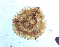
Dictyotriletes fimbriatus (Winslow) Kaiser, 1970
Radial, trilete, acamerate miospores. Amb rounded triangular to subcircular. Trilete mark distinct or obscured by ornamentation, sutures simple, straight, extending between half and two-thirds of spore radius. Exine bears characteristic reticulate ornament, muri present on proximal surface, reduced in height and density but commonly developed parallel to sutures of trilete mark. Muri 8–20 µm high and approximately 3 µm wide at base, with distinctive papillate crests. Muri enclose a variable number of large lumina up to 30 µm in diameter, variable in shape. Crests of muri deeply incised to about half their height to form blunt, slightly pointed or clavate papillae.
Winslow, 1962, pp. 58–59, pl. 14, figs. 1–3, pl. 22, fig. 21. Kaiser, 1970, p. 95, pl. 19, figs. 4–6
Overall equatorial diameter 69–115 µm. Diameter excluding ornament 52–80 µm
The distinctive papillate crested muri forming the reticulate ornamentation of this species render it readily distinct from all other taxa assigned to this genus.
Late Devonian (Strunian) to Mississsippian; Tournaisian; LL to HD Biozones (Higgs et al., 1988).
Dictyotriletes muricatus (Kosanke) Smith & Butterworth, 1967
Radial, trilete, acamerate miospores. Amb circular to rounded triangular. Trilete mark distinct or obscured by the ornamentation, sutures simple straight, extending between half and three-quarters of spore radius. Exine ornamented on entire distal surface and equatorial portion of proximal surface with muri which are up to 2 µm wide and 6–12 µm high. Muri enclose variable number of polygonal lumina which are of more or less uniform size on any individual but vary in size and number between individuals. Lumina vary in diameter up to 25 µm. Exine of lumina laevigate although infrequent small coni and spinae may occur.
Kosanke, 1950, p. 27, pl. 4, fig. 7. Smith and Butterworth, 1967, pp. 197–198, pl. 11, figs. 25–26
Overall equatorial diameter 68(83)97 µm
Some specimens assigned to this species may possess lumina which are closely comparable in size and form to those of Reticulatisporites reticulatus (Ibrahim) Ibrahim 1933. The latter species is however readily distinguished by possessing a differentially thickened cingulum.
Pennsylvanian; Bashkirian to Moscovian [Langsettian to early Bolsovian] (Smith and Butterworth, 1967)
Endosporites zonalis (Loose) Knox, 1950
Radial, trilete, camerate miospores. Amb of body and saccus circular to rounded triangular. Trilete mark distinct, sutures straight, extending to or almost to margin of inner body, accompanied by elevated flexuous folds of the exoexine which extend, decreasing in height, almost to the equatorial margin of the exoexinal body. Ends of elevated exoexinal folds connected laterally with narrow, fine, curvatural ridges that in proximo-distal compression are located, for much of their length coincident with the equatorial margin of the saccus, the exception being in the radial positions where minor invagination takes place. Intexine thinner than exoexine, forms inner body that may possess peripheral taper-point compression folds, indicating significant separation of the two exine layers. Exoexine thin, granulate with fine narrow limbus, forms saccus and is commonly extensively folded.
Loose, 1934, p. 148, p. 17, fig. 5. Smith and Butterworth, 1967, pp. 272–273, pl. 22, figs. 3–4
Overall equatorial diameter 61(82)104 µm. Diameter of inner body 33(50)61 µm. Ratio of inner body to saccus diameter 46(59)77 µm
Almost identical to Endosporites globiformis (Ibrahim) Schopf, Wilson & Bentall except the ratio of the inner body to saccus is consistently larger and the exoexine appears slightly thicker.
Pennsylvanian; Bashkirian to Moscovian [late Langsettian to Asturian] (Smith and Butterworth, 1967).
Grandispora lupata Turnau, 1975
Radial, trilete camerate miospores. Amb rounded triangular with convex sides and rounded apices. Trilete mark distinct, sutures straight, accompanied by elevated flexuous folds of the exoexine which extend, decreasing in height, towards the equatorial margin of spore. Exoexine separated from intexine at least in the equatorial region and over part of distal surface. Intexine thin, forming rounded triangular inner body occupying up to three-quarters of the total spore diameter. Exoexine finely infrapunctate, relatively thick over distal surface and equatorial portion of proximal surface, thinner over contact areas which occupy approximately two-thirds of spore radius. Ornamentation of exoexine confined to the distal surface and consists of spinae with bulbous bases and sharply pointed terminations. Elements up to 2.5 µm high and 1.5 µm in basal diameter, varying between 1.5 and 9 µm (commonly 5 µm) apart.
Turnau, 1975, pp. 517–518, pl. 6, figs. 1–3
Overall equatorial diameter 64(75)95 µm
Grandispora echinata Hacquebard 1957, is distinguished by possessing a thinner exoexine which is of uniform thickness, and a more obvious intexine forming the inner body. Grandispora gracilis (Kedo) Streel in Becker et al. 1974 is smaller, has a thinner exoexine and possesses a finer ornamentation. Grandispora cf. uncata (Hacquebard) Playford 1971, is smaller and has a relatively coarser but less densely distributed ornamentation.
Devonian; Famennian (from Higgs et al., 2000)
Grandispora spinosa Hoffmeister, Staplin & Malloy, 1955
Radial, trilete, camerate miospores. Amb circular, subcircular or rounded triangular, outline of inner body more or less conformable and occupying two-thirds to four fifths of the spore diameter. Trilete mark distinct or obscured by folding, sutures straight, extending at least three-quarters of the inner body radius, commonly accompanied by elevated flexuous folds of the exoexine that extend decreasing in height to the equatorial margin of spore. Intexine thin, laevigate, forms well-defined inner body with independent compression folds indicating major separation of the two exine layers. Exoexinal body wall 1–2.5 µm thick; exoexine infrapunctate, ornamented over entire distal surface and equatorial portion of proximal surface with sharply pointed coni and spinae, many of which may have bulbous bases. Elements with bulbous bases more commonly located in the polar regions with simple, sharply-pointed coni and spinae at the equator. Elements 2–8 µm high (commonly 2–5 µm) and 2–4 µm in basal diameter. Exoexine commonly folded due to compression.
Hoffmeister, Staplin and Malloy, 1955, pp. 388–389, pl. 39, figs. 10 and 14. Sullivan and Marshall, 1966, p. 280, pl. 4, fig. 5
Overall equatorial diameter 80(105)165 µm. Diameter of inner body 62(89)107 µm
Distinguished from Grandispora echinata Hacquebard 1957, by its larger size and ornament of more widely spaced, bulbous-based spinae and coni.
Mississippian; late Visean to Serpukhovian [Arnsbergian]; VF to SO biozones (Clayton et al., 1977; Turner et al., 1994)
Grumosisporites varioreticulatus (Neves) Smith & Butterworth, 1967
Radial, trilete, camerate miospores. Amb subcircular to oval. Trilete mark distinct, sometimes obscured, sutures straight, simple or with low, broad, thickened labra, extending between two-thirds and three-quarters of the spore radius. Intexine thin, forming irregular to sub-triangular inner body. Exoexine 2–3.5 µm thick, ornamented with often poorly defined reticulate sculpture. Muri narrow, up to 2 µm high enclosing lumina of irregular shape which may be up to 15 µm in diameter. Muri project at equatorial margin as low, broad-based conate projections. Reticulum best developed on distal surface and is most apparent at slightly elevated levels of focus.
Neves, 1961, p. 8, pl. 2, figs. la–b. Smith and Butterworth, 1967, p. 232, pl. 17, figs. 8–10
Overall equatorial diameter 70–110 µm
The poorly defined reticulate ornament on the exoexine distinguishes G. varioreticulatus from other species assigned to the genus.
Pennsylvanian; Bashkirian to Moscovian [Bolsovian]; KV to SL biozones (Clayton et al., 1977).
Grumosisporites verrucosus (Butterworth & Williams) Smith & Butterworth, 1967
Radial, trilete, camerate miospores. Amb circular to rounded-triangular with undulating equatorial margin. Trilete mark distinct, sutures straight, simple or with low, narrow, thickened labra; extending between two-thirds and the full spore radius. Intexine thin, forms irregular, extensively folded, inner body which is separated from the exoexine over a significant part of its surface. The two layers appear to be in contact at least at the contact areas. Exoexine 1–4 µm in thickness, ornamented with variably shaped verrucae which show basal coalescence to form short, irregular ridges. Elements densely distributed and separated by narrow channels of unornamented exoexine. Up to 30 elements project at the equator to produce undulating margin. Verrucae up to 3 µm high at equator. Ornament reduced or absent on the proximal surface.
Butterworth and Williams, 1958, p. 369, pl. 2, figs. 2–3. Smith and Butterworth, 1967, pp. 232–233, pl. 18, figs. 1–6
Overall equatorial diameter 42(53)64 µm
Grumosisporites rufus (Butterworth & Williams) Smith & Butterworth 1967 possesses a thicker exoexine and a less regular ornamentation. Camptotriletes corrugatus (Ibrahim) Potonié & Kremp 1956 has a coarser ornament and lacks evidence of separation of the exine layers.
Mississippian; late Visean to Serpukhovian [Arnsbergian]; VF to NC biozones (Clayton et al., 1977).
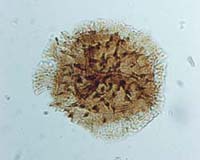
Hystricosporites multifurcatus (Winslow) Mortimer & Chaloner, 1967
Radial, trilete, acamerate miospores-megaspores. Amb, excluding projecting ornament, rounded triangular with straight or slightly convex sides and rounded apices. Proximal surface flattened, low, pyramidal; distal surface inflated, hemispherical. Trilete mark distinct, sutures straight, extending at least four-fifths of spore radius, accompanied by elevated flexuous folds of the exoexine which form an apical prominence up to 50 µm high. Folds decrease in height toward the equator. No obvious indication of separation of exine layers. Exine thick, surface scabrate, ornamented with long, parallel-sided or gently tapering processes which have expanded bases. Fusion between adjacent processes may occur at their bases and in the lower parts of the processes. Shafts of the processes appear hollow and have finely striated surfaces. Process termini are solid and characteristically ornamented with up to five recurved spinae producing a grapnel-like appearance. Processes 70–120 µm long, and excluding the expanded base, up to 10 µm wide. Recurved spinae creating grapnel terminations 1–5 µm long and 1–1.5 µm wide. Processes appear to be developed over entire distal surface with concentration in the equatorial region; 8–15 elements project at the equatorial margin. Some specimens show weak indication of ill-defined internal body.
Winslow, 1962, p. 52, pl. 11, figs. 4–5; p1. 12, fig. 5, p1. 22, fig. 15
Overall equatorial diameter excluding projecting ornament 90(173)233 µm
The distinctive ‘grapnel’ termini of its long slender processes distinguishes H. multifurcatus from all other taxa assigned to the genus. Mid and Late Devonian species of Hystricosporites have characteristic radially orientated ribs within the contact areas of the proximal surface; these are not present in H. multifurcatus.
Devonian to Mississippian; Strunian to early Visean (from unpublished information).
Knoxisporites hederatus (Ishchenko) Playford, 1962
Radial, trilete, cingulate miospores. Amb circular to subcircular, consisting of circular to subcircular spore body surrounded by more or less conformable cingulum. Trilete mark distinct, sutures simple, or accompanied by low, thickened labra up to 8 µm wide, which extend to the margin of the spore body. Sutures straight, extending between three-quarters to almost the full radius of the spore body. Exoexine extended in the equatorial plane to form undifferentiated cingulum, 6–16 µm in width. Distal surface bears conspicuous, widely spaced, relatively low, broad, smooth, rounded muri that are connected in several places to each other and to the cingulum. Muri slightly sinuous, irregular in pattern, 8–17 µm wide and 4–7 µm high. Remainder of exine laevigate.
Ishchenko, 1956, pp. 58–59, pl. 10, fig. 121 . Playford, 1962, pp. 634–635, pl. 90, figs. 9–12
Overall equatorial diameter 67(86)112 µm
The irregular development of low, broad muri on the distal surface distinguishes this species from other taxa assigned to Knoxisporites. Playford (1963) made no mention of the presence of low broad labra accompanying the trilete sutures that are a common feature of the Mattson Formation specimen illustrated here.
Mississippian; Tournaisian to Visean (Playford, 1962)
Knoxisporites stephanephorus Love, 1960
Radial, trilete, cingulate miospores. Amb circular, subcircular to rounded triangular. Trilete mark distinct, sutures straight, extending to margin of spore body, simple or accompanied by low narrow thickened labra which typically become expanded equatorially to form triangular thickened areas that fuse to the proximal surface of the cingulum. Exine laevigate. Intexine forming spore body rarely discernible. Exoexine closely adpressed to intexine but extended in the equatorial plane to form thickened, undifferentiated cingulum, 5–15 µm wide. Exoexine developed into second ring of thickening mid way between the equator and the distal pole with irregular, radially-orientated thickened ribs in the inter-radial positions sometimes connecting it to the cingulum. A distal polar thickening 5–10 µm in diameter is also developed on the exoexine.
Love, 1960, pp. 118–119, pl. 2, figs. 1–2
Overall equatorial diameter 40–90 µm
Knoxisporites rotatus Hoffmeister, Staplin & Malloy 1955 is distinguished by the absence of the distal polar thickening and by the presence of more prominent, radially-orientated ribs connecting the cingulum to the distal, circular ring of thickening.
Mississippian to Pennsylvanian; Visean to Moscovian [Asturian]; TS to OT biozones (Clayton, 1985, and unpublished information)
Knoxisporites triangularis Higgs, Clayton & Keegan, 1988
Radial, trilete, cingulate miospores. Amb subcircular to rounded triangular. Trilete mark distinct, sutures straight, sometimes open, extending to the inner margin of the cingulum, accompanied by broad prominent labra up to 20 µm in overall width. Intexine relatively thin; forms rounded triangular to subcircular inner body. Exoexine extended in equatorial plane to form undifferentiated cingulum, up to 14 µm wide. Proximal surface laevigate. Distal surface ornamented with three muri arranged in a triangular pattern with the apices of the triangle joining the cingulum in the inter-radial positions. Muri vary between 8–12 µm in width and are often slightly expanded where they fuse with the cingulum. Remainder of exine laevigate to scabrate.
Higgs, Clayton and Keegan, 1988, p. 66, pl. 8, figs. 3,6
Overall equatorial diameter 79(91)121 µm
The distinctive triangular structure created from three muri on the distal surface distinguish this species from others assigned to the genus Knoxisporites. Higgs, Clayton and Keegan (1988) elevated this form to specific status, considering that it was sufficiently distinct from Knoxisporites literatus (Waltz) Playford 1963 within which it had previously been allocated varietal status.
Mississippian; Tournaisian; Tn1b to Tn3c, VI to CM biozones (Higgs et al., 1988)
Knoxisporites triradiatus Hoffmeister, Staplin & Malloy, 1955
Radial, trilete, acamerate miospores. Amb subcircular, sometimes rounded triangular. Trilete mark distinct, sutures straight, extending to, or almost to, the equatorial margin of the spore body; sutures accompanied by low, broad, thickened labra. Intexine probably thin; forms inner spore body conformable in outline with the equatorial margin. Exoexine closely adpressed to intexine over proximal and distal surfaces but extended in the equatorial plane to form thick undifferentiated relatively narrow cingulum. Distal surface of exoexine bears thickened, Y-shaped structure with ribs of uniform thickness, extending from the distal pole to fuse with the cingulum in the inter-radial positions.
Hoffmeister, Staplin & Malloy, 1955, p. 391, pl. 37, figs. 11–12
Overall equatorial diameter 50–88 µm
The regular form of the Y-shaped thickening on the distal surface, and the narrow undifferentiated cingulum distinguish this species from others assigned to the genus Knoxisporites. Knoxisporites seniradiatus appears to have thickenings associated with the proximal trilete mark.
Mississippian; Visean to Serpukhovian [Arnsbergian]; TS to SO biozones (Clayton, 1985, and unpublished information)
Kraeuselisporites echinatus Owens, Mishell & Marshall, 1976
Radial, trilete, camerate, zonate miospores. Amb rounded triangular to subcircular. In lateral compression proximal surface low, pyramidal; distal surface hemispherical. Trilete mark distinct, sutures straight, extending to margin of inner body, accompanied and obscured by elevated, flexuous folds of the exoexine, up to 8 µm high in the polar region but decreasing in height to the equatorial margin. Intexine thin, forming rounded triangular to subcircular inner body. Exoexine extended in the equatorial plane to produce strongly bizonate zona up to 25 µm wide. Exoexine thicker over distal surface of inner body and extending into the inner part of the zona, outer part of zona thinner and weakly striated. Two exine layers normally closely adpressed but minor separation may occur in the equatorial plane and over the more equatorial portions of the distal surface. Exoexine surface finely infrapunctate, distal surface bears distinctive ornament of large pointed coni and spinae with expanded bases; height of elements 4–14 µm, basal diameter 2–7 µm. Elements variable in distribution with maximum concentration usually occurring on the inner thicker zone of the zona close to the margin of the spore body.
Owens, Mishell and Marshall, 1976, pp. 148–153, pl. 1, figs. 1-6, pl. 2, fig. 1
Overall equatorial diameter 74(104)147 µm. Maximum equatorial diameter of inner body 54(71)82 µm
Kraeuselisporites ornatus (Neves) Owens, Mishell & Marshall 1976, differs by possessing an ornament of apparently smaller, delicate coni and spinae. Kraeuselisporites scorpius Balme & Hassell 1962, described from the Upper Devonian of the Canning Basin, Western Australia is distinguished by possessing a narrower flange and a more densely distributed ornament of shorter, stouter, broad-based coni, spinae and irregular baculae.
Mississippian to Pennsylvanian; late Visean to Bashkirian [Marsdenian]; VF to KV biozones (Clayton et al., 1977, and unpublished information)
Kraeuselisporites hibernicus Higgs, 1975
Radial, trilete, camerate miospores. Amb subcircular to rounded triangular with convex sides and rounded or slightly pointed apices. Trilete mark distinct or obscure; sutures straight or sinuous, extending to margin of spore body, accompanied by elevated flexuous folds of the exoexine which extend, decreasing in height, to the equatorial margin of the spore. Intexine thin, forming subcircular to rounded triangular inner body. Exoexine slightly thicker over distal surface and extended in the equatorial plane to form slightly fibrous zona 8–18 µm in width. Distal and more equatorial portions of proximal surface of exoexine ornamented with sparse to closely spaced spini, coni and grana. Elements either discrete or fused at their bases to form short irregular cristae or rugulae. Spinae 1–5 µm high and 0.5–2 µm in basal diameter, coni 1–3 µm in height and grana up to 1 µm high. Exoexine of proximal surface thin and crumpled and in many cases folded along the line of the margin of the intexine.
Higgs, 1975, p. 400, pl. 6, figs. 11–12. Higgs, Clayton and Keegan, 1988, p. 79, p1. 16, fig. 3
Overall equatorial diameter 64(85)116 µm. Diameter of the inner body 40(53) 70 µm
Mississippian; Tournaisian; HD to CM biozones (Higgs et al., 1988)
Kraeuselisporites ornatus (Neves) Owens, Mishell & Marshall, 1976
Radial, trilete, cingulizonate, camerate miospores. Amb rounded triangular to subcircular, in lateral profile proximal surface flattened, low or pyramidal; distal surface hemispherical. Trilete mark distinct, sutures straight, extending to, or almost to, the equatorial margin of spore body, accompanied by elevated flexuous folds of the exoexine which extend decreasing in height to the margin. Exine composed of two layers. Intexine, thin, forms rounded triangular to subcircular inner body. Exoexine normally closely adpressed to the intexine but displays limited separation in the equatorial region. Exoexine extends to form bizonate cingulum. Maximum thickness of the exoexine developed over the distal surface and in the region of the cingulum adjacent to inner body margin. Outer zone of cingulum thinner and wider; sometimes appears weakly striated. Distal surface of exoexine ornamented with sharply pointed coni and spinae. Elements 2–10 µm high (commonly 3–5 µm) with the longest most slender elements located close to the junction of the body and cingulum.
Neves, 1961, p. 269, pl. 33, fig. 3. Owens, Mishell and Marshall, 1976, pp. 153–154, pl. 2, figs. 2–4
Overall equatorial diameter 76–110 µm
Kraeuselisporites ornatus is distinguished from K. echinatus Owens, Mishell & Marshall 1976, by its smaller overall size and its proportionately smaller ornament of shorter, more slender coni and spinae.
Mississippian to Pennsylvanian; Serpukhovian to Bashkirian [Arnsbergian to Langsettian]; SO to RA biozones (Clayton et al., 1977)
Labiadensites fimbriatus (Waltz) Hacquebard & Barss, 1957
Radial, trilete, cingulate miospores. Amb circular to subcircular. Trilete mark distinct, sutures straight, extending almost to spore body margin, accompanied by broad, flat labra, extending 6–10 µm on either side of the sutures and having undulate outer margins. Intexine laevigate to infrapunctate, forms circular to subcircular inner body. Exoexine extended in the equatorial plane to form smooth dense cingulum surrounded by more transparent, less robust, equatorial border which has a frilled membranous appearance. Boundary between the two parts of the cingulum is smooth. In some specimens outer membranous part of the cingulum appears to overlap thicker inner part particularly on the distal surface. Thinner outer membranous section may be variable in width creating undulate equatorial margin.
Hacquebard and Barss, 1957, p. 28, pl. 4, fig. 2 . Playford, 1962, pp. 632–633, pl. 90, figs. 1–3
Overall equatorial diameter 90(115)144 µm. Diameter of the inner body 50(69)88 µm
Playford (1963) pointed out that those specimens in which the thinner membranous zone of the cingulum has been partly removed appear as transitional forms with Anulatisporites labiatus Hughes & Playford 1961, which has a smooth undifferentiated cingulum.
Mississippian; Tournaisian to Visean (Playford, 1962)
Lophozonotriletes malevkensis Naumova in litt. Kedo, 1963
Radial, trilete, cingulate miospores. Amb rounded triangular to subcircular, inner body conformable in outline with the equatorial margin. Trilete mark distinct to indistinct, sutures simple, straight, extending to margin of the inner body, sometimes accompanied by low narrow labra. Intexine thin, laevigate, forms inner body conformable in outline with the equatorial margin. Exoexine closely adpressed to intexine over both proximal and distal surfaces but extended in the equatorial plane to form more or less undifferentiated uniform cingulum which occupies 20–35 % of the spore radius. Distal surface of exoexine ornamented in the region underlying the inner body with prominent rounded tubercles and verrucae, elements normally discrete but variable in size, height up to 6 µm and basal diameter varying between 3–15 µm. Elements commonly distributed in a concentric manner in the region adjacent to the body-cingulum boundary with a single, frequently larger, element developed in the distal polar region. Ornamentation absent from the majority of cingulum, margin smooth or minutely indented. Surface of exoexine between verrucose elements smooth or minutely granulate.
Kedo, 1963, p. 87, pl. 10, figs. 240–241. Turnau, 1978, p. 9, pl. 3, fig. 8. Higgs, Clayton and Keegan, 1988, p. 67, pl. 8, figs. 11–13
Overall equatorial diameter 32(42)52 µm
Playford (1991) considered L. malevkensis and L. rarituberculatus (Luber) Kedo to be synonymous and to be more appropriately accommodated in Tumulispora Staplin & Jansonius 1964. In the present work the two taxa are maintained and retained in Lophozonotriletes, which is considered to be adequately differentiated from Tumulispora. Lophozonotriletes malevkensis and L. rarituberculatus differ in the following ways. · L. malevkensis (32(42)52 µm; present study) is smaller than L. rarituberculatus (54(76)100 µm; present study). · The ornamentation of verrucae and tubercles is smaller in L. malevkensis. · The distribution of the verrucate ornament in L. malevkensis is restricted to the more polar regions of the distal surface and is largely absent from the cingulum resulting in a smooth equatorial margin. In L. rarituberculatus some of the ornament projects at the equator.
Late Devonian to Mississippian; Strunian to Tournaisian; LN–PC biozones (Clayton et al., 1977)
Lycospora pusilla (Ibrahim) Somers, 1972
Radial, trilete, cingulizonate miospores. Amb circular to very broadly rounded triangular, outline smooth or finely indented. Trilete mark distinct, sutures simple or accompanied by low narrow flexuous exoexinal folds which extend decreasing in height to equatorial margin. Sutures extend to inner body margin. Intexine thin forming rounded triangular inner body. Exoexine thin, extended in the equatorial plane to form narrow bizonate cingulum consisting of a narrow, inner, darker zone which may in part overlap the equatorial margin of the inner body and a wider, thinner, outer zone. Total width of cingulum up to 4 µm. Distal surface of exoexine and cingulum finely granulate or in some cases conate. Folding common.
Schopf, Wilson and Bentall, 1944, p. 54. Smith and Butterworth, 1967, pp. 251–252, pl. 20, figs. 10–12. Somers, 1972, pp. 66–70, pl. 1, figs. 5–33, pl. 2, figs. 3–20
Overall equatorial diameter 20(27)40 µm
Lycospora noctuina Butterworth & Williams 1958, is distinguished from L. pusilla by the presence of rugulae or verrucae on the distal surface. L. orbicula (Potonié & Kremp) Smith & Butterworth 1967, differs in possessing a very narrow cingulum. (See Somers, 1972, for detailed appraisal of L. pusilla and its relationships to other members of the genus.)
Mississippian to ?Permian; Visean to ?Asselian; Pu to Granulatisporites confluens biozones (various sources)
Monilospora moniloformis Hacquebard & Barss, 1957
Radial, trilete, cingulizonate miospores. Amb subcircular to rounded triangular, with straight to slightly concave or convex sides and rounded apices. Trilete mark distinct, sutures simple, straight; extending from two-thirds to almost the full body radius, frequently open. Intexine thin, laevigate, forming inner body. Exoexine normally closely adpressed to intexine, although minor separation may occur in the equatorial plane. Exoexine extended in the equatorial plane to form trizonate differentiated cingulum consisting of a broad, thickened inner zone of uniform thickness with a sharp outer margin, a thinner median zone which is commonly narrow, and a distinctive crenulate, outer, thickened zone which varies considerably in form and intensity of crenulation. Surface of exoexine laevigate.
Hacquebard and Barss, 1957, p. 38, pl. 5, figs. 8, 9
Overall equatorial diameter 58–86 µm
Mississippian, late Visean, [Asbian] NM Biozone (unpublished information)
Murospora parthenopia Neves & loannides, 1974
Radial, trilete, cingulate miospores. Amb rounded triangular with concave, rarely straight sides, and variably rounded apices. Spore body rounded triangular with concave sides and rounded apices. Trilete mark distinct, sutures simple, straight, sometimes open, extending to equatorial margin of spore body. Exoexine extended in the equatorial plane to produce equatorial cingulum of variable width, expanded in the apical regions to form thickened valvae, 3–15 µm high and 9–20 µm wide. Inner margin of thickening may transgress onto the spore body on the distal surface; outer margin smooth or indented owing to minor fluting. Inter-radially, cingulum is variable in form, sometimes the same width as valvae but not thickened; or alternatively narrow to almost absent. Distal surface of spore body may develop rudimentary thickened kyrtome-like structures. Exine laevigate.
Neves and loannides, 1974, pp. 77–78, pl. 6, figs. 14–15
Overall equatorial diameter 26(34)50 µm
Neves and loannides (1974) considered this species to be synonymous with specimens recorded as Triquitrites coesfeldens by Bharadwaj and Venkatachala (1961).
Mississippian; late Visean; NM Biozone (DP to ME sub-biozones) (Clayton et al., 1978)
Radiizonates aligerens (Knox) Staplin & Jansonius, 1964
Radial, trilete, cingulizonate miospores. Amb rounded triangular to subcircular. Trilete mark indistinct, sutures when seen, simple, straight or flexuous, extending to, or almost to, inner body margin. Intexine thin, laevigate, forming poorly defined inner body that may show evidence of minor separation from the exoexine in the equatorial plane. Exoexine extended in the equatorial plane to form weakly bizonate cingulum consisting of a narrow darker inner zone which appears to overlap the equatorial margin of the inner body, particularly over the distal surface, and a wider thinner outer zone. Outer zone of cingulum bears a variable number of radially orientated elongate vacuoles that appear to be independently developed at two levels, presumably underlying both proximal and distal surfaces. Exoexine surface also bears numerous radially orientated striations and plications, which extend to the equatorial margin. Vacuoles separated by narrow walls. Distal polar exoexine may bear scattered grana or small verrucae.
Knox, 1950, p. 329, pl. 19, fig. 288. Staplin and Jansonius, 1964, p. 106, p1. 18, figs. 23–28. Smith and Butterworth, 1967, p. 263, pl. 21, figs. 9–11
Overall equatorial diameter 47(60)85 µm. Width of cingulum 14(19)27 µm
Pennsylvanian; Bashkirian [Langsettian]; RA Biozone (Clayton et al., 1977)
Raistrickia clavata Hacquebard 1957 emend. Playford, 1964
Radial, trilete, acamerate miospores. Amb rounded to convexly triangular. Trilete mark distinct or obscured by ornamentation; sutures simple, straight, extending up to three-quarters of spore radius. Exine 2–3 µm thick and ornamented with conspicuous discrete, short, club-shaped, baculate processes. Elements 5–10 µm high, 2–4 µm in basal diameter with slightly tapering shafts before expanding into a bulbous or flattened termination 3–7 µm in width. Ornament predominantly developed on distal surface with minor elements on more equatorial portion of proximal surface.
Hacquebard, 1957, p. 310, pl. 1, fig. 25. Playford, 1964, pp. 24–25, pl. 6, figs. 5–10. Higgs, Clayton and Keegan, 1988, p. 56, pl. 4, fig. 13
Overall equatorial diameter 55(62)94 µm
Raistrickia ponderosa Playford 1964 is distinguished from R. clavata by being larger and possessing a thicker, more uniformly sculptured exine and a more constantly circular outline. R. strumosa Playford 1976 possesses a similar style of ornamentation but may differ in possessing more densely distributed processes and by having a thicker exine.
Mississippian; Tournaisian; PC to CM biozones (Higgs et al., 1988)
Raistrickia condylosa Higgs, 1975
Radial, trilete, acamerate, miospores. Amb circular-subcircular. Trilete mark distinct or obscured by ornament, sutures simple, straight, extending between half and two-thirds spore radius. Exine 1–3 µm thick, bearing prominent baculose ornament, which may be densely or sparsely distributed but reduced on the proximal surface. Elements with slender shaft, long, parallel sided, slightly curved; surmounted by rounded or more commonly bulbous termination. Pointed elements rare. Bases of individual elements normally discrete but may fuse laterally to form low ridges 1–3 µm in height. Elements 5–30 µm long, 1–4 µm in width, bases 3–7 µm in diameter and bulbous terminations up to 6 µm wide.
Higgs, 1975, p. 396, pl. 2, figs. 15–16. Higgs, Clayton and Keegan, 1988, p. 56, pl. 4, figs. 15–16
Equatorial diameter excluding projecting ornament 66(87)120 µm
Raistrickia strumosa Playford 1976 has a similar style of ornament to R. condylosa but its elements are more robust and more densely distributed. Acanthotriletes halei Varma 1969 appears superficially similar but may be distinguished by possessing longer sutures and pointed ornamentation elements.
Mississippian; Tournaisian; PC to CM biozones (unpublished information)
Raistrickia corynoges Sullivan, 1968
Radial, trilete, acamerate miospores. Amb subcircular. Trilete mark distinct to obscured, sutures simple or with narrow thickened labra, extending at least three-quarters of spore radius. Exine 2–3 µm thick bearing dense ornament of prominent bacula. Bases of individual elements wide and sometimes coalescent. Shafts are commonly parallel sided, but may taper slightly, sometimes with terminal constrictions. Terminations truncated, rounded or pointed, rarely weakly multifurcate. Length of elements 8–26 µm, width 2–7 µm.
Sullivan, 1968, p. 119–120, p1. 25, figs. 6–8. Higgs, Clayton and Keegan, 1988, p. 58, pl. 4, fig. 11
Equatorial diameter excluding projecting ornament 60(73)95 µm
Raistrickia variabilis Neves & Dolby 1970 differs by possessing more robust, more densely distributed bacula that are often fused at their bases. R. macrura (Luber) Dolby & Neves 1970 is distinguished by possessing bacula which are smaller and more tapered in profile.
Mississippian; Tournaisian; VI to CM biozones (Higgs et al., 1988)
Raistrickia fulva Artüz, 1957
Radial, trilete, acamerate miospores. Amb rounded triangular. Trilete mark distinct, sutures simple, straight, extending two-thirds to three-quarters of spore radius. Distal surface and equatorial portion of proximal surface of exine bears predominantly baculate ornament with infrequent coni and verrucae. Baculae rarely exceed 3 µm in height, basal diameter exceptionally up to 10 µm but commonly less, terminations truncate or apiculate. Density of ornament variable with 15–20 processes commonly protruding at the equator. Exine between processes laevigate; up to 5 µm thick.
Artüz, 1957, p. 246, pl. 3, fig. 19. Smith and Butterworth, 1967, pp.180–181, pl. 8, figs. 17–20
Overall equatorial diameter 39(52)69 µm
Raistrickia irregularis Kosanke 1950 is larger. No other species assigned to the genus Raistrickia has such low, broad-based baculae.
Pennsylvanian; Bashkirian [Kinderscoutian to Duckmantian]; KV to NJ biozones (Clayton et al., 1977)
Reinschospora speciosa (Loose) Schopf, Wilson & Bentall, 1944
Radial, trilete, miospores. Amb including corona rounded triangular to circular. Outline of spore body triangular with rounded apices and concave sides. Trilete mark distinct, sutures simple or with weak narrow low labra; straight, extending almost to equatorial margin. Well-developed corona composed of setae up to 1 µm wide and more or less in full lateral contact throughout their length. Corona attached to exine just proximally of the equatorial margin. Maximum width of corona, including overlap on proximal side, 17–28 µm; occurs in the inter-radial position. In the radial positions the corona may be as narrow as 2 µm and may be attached nearer to the proximal pole so that on compression it may appear to cross the angles of the spore body. Exine moderately thick, laevigate to scabrate, folding rare.
Schopf, Wilson and Bentall, 1944, p. 53, fig. 2. Smith and Butterworth, 1967, pp. 211–212, pl. 13, figs. 13–14
Equatorial diameter of spore body 50(58)95 µm
Reinschospora triangularis Kosanke 1950 is distinguished from R. speciosa in possessing straight sides, labra, and setae which are discrete and commonly partate. R. magnifica Kosanke 1950, differs by having broader angles to the spore body and a corona composed of discrete elements.
Pennsylvanian; Bashkirian to Moscovian [Kinderscoutian to Bolsovian] (Smith and Butterworth, 1967, and unpublished information)
Remysporites magnificus (Horst) Butterworth & Williams, 1958
Radial trilete camerate miospores. Amb ovoid to circular. Margin smooth to minutely undulating. Trilete mark distinct or obscured by elevated exoexinal folds which extend, decreasing in height, to equatorial margin. Sutures, when seen, extend almost to inner body margin. Intexine relatively thick, laevigate, with independent compression folds indicating complete separation from exoexine over most of its surface. Exoexine thin, forms highly folded exoexinal body; surface laevigate to microreticulate. Exoexine of contact areas may develop fine vermiculate ornament.
Horst, 1955, p. 194, pl. 21, fig. 37. Butterworth and Williams, 1958, p. 386, pl. 4, figs. 7–9. Smith and Butterworth, 1967, p. 278, pl. 33, fig. 8
Overall equatorial diameter 84–255 µm
Smith and Butterworth (1967) considered that Remysporites albertensis Staplin 1960 is synonymous with the present species.
Mississipian; late Visean to Serpukhovian [Asbian to Arnsbergian] (Smith and Butterworth, 1967, and unpublished information)
Reticulatisporites carnosus (Knox) Neves, 1964
Radial, trilete, cingulate miospores. Amb broadly rounded triangular to subcircular, margin usually smooth, but occasionally undulate to uneven or highly involute. Trilete mark distinct, sutures straight, simple or with low narrow labra, extending to inner margin of the cingulum. Intexine relatively thin forming subcircular to rounded triangular inner body. Exoexine extended equatorially to form differentially thickened cingulum consisting of three zones, each varying from 5 to 17 µm in width. The inner thickened zone is broader and more prominent than the outer marginal thickened zone, which is commonly reduced or discontinuous. The thinner median zone may be reduced to a mere line but in other specimens may be of significant width. Radially orientated thickenings may be present on the distal surface and take the form of a Y-shaped structure attached to the cingulum in the inter-radial positions. Faint proximal thickenings may arise from the radial positions but they scarcely extend poleward beyond the inner thickened zone of the cingulum.
Knox, 1950, p. 329, p1. 19, fig. 290. Smith and Butterworth, 1967, pp. 220–221, pl. 14, figs. 11–12
Overall equatorial diameter 67(84)90 µm
Distinguished from other species assigned to the genus by the absence of any elaborate pattern of distal thickenings. In some specimens assigned to this species a distal pattern of thickenings comparable to those in Reticulatisporites polygonalis (Ibrahim) Neves 1964 may occur, in which case R. carnosus is distinguished by the discontinuous nature of the outermost thickened zone of its cingulum.
Mississippian to Pennsylvanian; late Visean to Bashkirian [early Langsettian]; NC to SS biozones (Clayton et al., 1977; Smith and Butterworth, 1967)
Retispora lepidophyta (Kedo) Playford, 1976
Radial, trilete, camerate miospores. Amb convexly rounded triangular to subcircular. Trilete mark distinct; sutures straight or slightly curved, extending to margin of the inner body, accompanied by elevated flexuous folds of the exoexine which extend decreasing in height to almost the equatorial margin of the spore. Ends of exoexinal folds fuse laterally with curvaturae imperfectae, which are slightly invaginated in the radial position but are coincident with the equatorial margin for much of their length. Intexine thin; forms subcircular to rounded triangular inner body which occupies half to three-quarters of the spore diameter. Sometimes eccentrically placed, suggesting proximal attachment only. Exoexine ornamented with distal reticulum consisting of subcircular, polygonal or irregularly shaped lumina separated by narrow muri. Muri 0.5–2 µm wide; bear an ornament of small coni and spinae up to 2 µm high. Muri commonly exhibit progressive breakdown to form short rugulae or eventually residual coni and verrucae. Lumina up to 4 µm in diameter, variable in shape and size even on same specimen. Narrow limbus may be present at the equatorial margin, which may in part coincide with curvaturae imperfectae. Apical papillae sometimes present. Compressional folds common.
Kedo, 1957, p. 24, pl. 2, figs. 19–21. Streel in Becker et al., 1974, p. 26. Playford, 1976, p. 45, pl. 10, figs. 1–15. Higgs, Clayton and Keegan, 1988, pp. 71–72, pl. 11, figs. 12–17
Overall equatorial diameter 40(57)92 µm
The following taxa may be synonymous with Retispora lepidophyta: Leiozonotriletes naumovae Balme & Hassell 1962, Endosporites lacunosus Winslow 1962, Archaeozonotriletes fenestratus Caro-Moniez 1962 and Hymenozonotriletes reticulatus Caro-Moniez 1962.
Devonian; late Famennian to early Tournaisian; LL–LN biozones (Richardson and McGregor, 1986; Higgs et al., 1988)
Rotaspora fracta Schemel, 1950
Radial, trilete acamerate miospores. Amb circular, subcircular or rounded triangular, spore body triangular with slightly concave sides and rounded apices. Trilete mark distinct, sutures straight, simple or accompanied by low narrow labra, extending almost to spore body margin. Width of zona more or less constant, with narrow thickening at the periphery forming equatorial rim. In compressed specimens the zona lies over the distal surface of the body in the radial positions thereby appearing narrower than in the inter-radial areas. Exine of body and zona laevigate. Folding rare.
Schemel, 1950, p. 242, pl. 40, fig. 8. Smith and Butterworth, 1967, p. 227, pl. 15, figs. 8–11
Overall equatorial diameter 24–40 µm. Diameter of spore body 17–24 µm
Distinguished from Rotaspora knoxi Butterworth and Williams 1958, by its slightly smaller size, thinner exine and spore body with concave sides.
Mississippian; late Visean to Serpukhovian [Pendleian]; VF to NC biozones (Clayton et al., 1977, and unpublished information)
Rotaspora knoxi Butterworth & Williams, 1958
Radial, trilete, acamerate miospores. Amb subtriangular, with broadly rounded apices, to subcircular, outline of body more triangular. Trilete mark distinct; sutures straight, simple or with low narrow labra, extending between three-quarters and almost the full radius of the body. Exine laevigate. Zona widest (up to 6 µm) in the inter-radial position but significantly reduced at the apices. Zona normally bizonate with inner thicker zone of more or less uniform width, apparently overlapping margin of inner body at least on the distal surface. Outer thinner zone variable in width.
Butterworth and Williams, 1958, p. 378, pl. 3, figs. 21–23. Sullivan and Marshall, 1966, p. 272, pl. 2, figs. 16–17. Smith and Butterworth, 1967, p. 228, pl. 15, figs. 15–17
Overall equatorial diameter 26(32)44 µm
Rotaspora fracta Schemel 1950, is distinguished by its smaller size, its concavely triangular spore body, its thinner exine and the tendency of its zona to be folded over the body in the radial positions.
Mississipian; late Visean to Serpukhovian [Arnsbergian]; VF to TK biozones (Clayton et al., 1977).
Schopfites claviger Sullivan, 1968
Radial, trilete, acamerate or camerate miospores. Amb subcircular or circular. Trilete mark distinct or indistinct, sutures simple, straight, extending almost to the equatorial margin. Intexine thin (1–1.5 µm), indistinctly separated from the exoexine to form indistinct inner body occupying approximately three-quarters of spore diameter. Distal surface and equatorial region of exoexine ornamented with pila, rounded bacula and rare verrucae and coni. Elements 1–5 µm high and 1–3 µm in width (Higgs et al., 1988, quoted mean height and width as 3 µm and 1 µm respectively). Elements are normally discrete, although the density and distribution of the ornament is variable, giving rise to a patchy appearance on individual specimens. Proximal surface of exoexine laevigate. Exine thickness 1–2 µm.
Sullivan, 1968, p. 121, pl. 25, figs. 9–10. Higgs, Clayton and Keegan, 1988, pp. 59–60, pl. 5, figs. 6–8 and 13
Overall equatorial diameter 40(55)68 µm
Although complete morphological intergradation is thought to exist between Schopfites claviger and S. delicatus Higgs 1975, the latter species is distinguished by possessing an ornament of finer sculptural elements that does not include rare coni and verrucae.
Mississippian; late Tournaisian to early Visean; CM–Pu biozones (Higgs et al., 1988).
Schulzospora campyloptera (Waltz) Hoffmeister, Staplin & Malloy, 1955
Radial, trilete, camerate miospores with a bilaterally symmetrical appearance due to the presence of an elliptical saccus-like exoexinal structure. Amb oval but occasionally sub-circular. Trilete mark distinct to indistinct; sutures straight, extending up to two-thirds of spore body radius, sutures often of unequal length on individual specimens. Intexine forms circular to oval inner body; long axis of inner body rarely orientated obliquely to the long axis of the miospore. Exoexine infrapunctate with a narrow dark zone often occurring where the saccus overlies the equatorial margin of the spore body. Radial compression folds infrequently developed.
Sullivan and Marshall, 1966, pp. 274–275, pl. 3, figs. 13–14. Smith and Butterworth, 1967, pp. 274–275, pl. 23, fig. 1
Long axis dimension 76(94)114 µm. Diameter of inner body 46(54)70 µm
Schulzospora rara Kosanke 1950 is distinguished from S. campyloptera by its more rounded equatorial outline and its proportionately larger spore body. The long axis of the inner body of S. campyloptera is not normally orientated obliquely to the long axis of the miospore, a characteristic which is typical of S. ocellata (Horst) Potonié & Kremp 1956. Intermediate forms may occur but it is not known if these represent morphological intergradations or are the result of compressional effects.
Mississipian; Visean to Serpukhovian [Arnsbergian]; TC to SO biozones (Clayton et al. 1977, and Varker et al., 1990)
Schulzospora ocellata (Horst) Potonié & Kremp, 1956
Radial, trilete, camerate miospores with a bilateral appearance due to the presence of an elliptical saccus-like exoexinal structure. Amb oval to subcircular, body oval to subcircular. Trilete mark distinct to indistinct; sutures simple, straight, extending between two-thirds and almost the full body radius. Exoexine infrapunctate with a narrow dark zone often occurring where the saccus overlies the equatorial margin of the spore body. Longest axis of the inner body usually oblique to the long axis of the miospore, rarely at right angles to it. Folding infrequent.
Horst, 1955, p. 195, p1. 21, figs. 40a and b. Smith and Butterworth, 1967, pp. 275–276, pl. 23, fig. 6
Overall equatorial diameter 61(90)130 µm. Diameter of inner body 56(66)77 µm
Smith and Butterworth (1967) considered this to be the largest of the species of Schulzospora. The specimens illustrated here fall at the smaller end of the size range quoted by them. The characteristic used to distinguish the species is the obliquity of the inner body within the saccus-like exoexinal structure. Morphological intergradation, is however, known to occur with Schulzospora campyloptera (Waltz) Hoffmeister, Staplin & Malloy 1955. Schulzospora rara Kosanke 1950 has a rounder, less elongated overall outline than Schulzospora ocellata.
Mississippian; late Visean to Serpukhovian [Arnsbergian]; NC to SO biozones (unpublished information, and Varker et al., 1990)
Schulzospora rara Kosanke, 1950
Radial, trilete, camerate miospores with a bilaterally symmetrical appearance due to presence elliptical saccus-like exoexinal structure. Amb oval to ovoid. Inner body circular to oval with long axis parallel to the long axis of the miospore overall. Trilete mark not always visible; sutures simple, straight extend to, or almost to, inner body margin. Two of the sutures are commonly orientated parallel or nearly parallel to the long axis of the miospore with the third suture at right angles to them. Intexine of inner body relatively thin, laevigate or finely infrapunctate; long axis of inner body approximately three-quarters of miospore long axis, short axis of inner body almost equal in width to the short axis of the miospore. Exoexine thicker than intexine, laevigate to finely infrapunctate.
Kosanke, 1950, p. 53–54, pl. 13, figs. 5–8. Smith and Butterworth, 1967, pp. 276–277, pl. 23, figs. 2–3
Overall equatorial diameter 52(77)105 µm. Diameter of inner body 40(59)80 µm
Schulzospora rara is similar in size to S. campyloptera (Waltz) Hoffmeister, Staplin & Malloy 1955, but may be distinguished by its more rounded, less elliptical shape and the proportionately larger size of the spore body.
Mississippian to Pennsylvanian; Visean to Bashkirian [Langsettian] (Smith and Butterworth, 1967)
Spelaeotriletes arenaceus Neves & Owens, 1966
Radial, trilete, camerate miospores. Amb convexly triangular, or subcircular to oval when secondarily folded. Trilete mark distinct; sutures simple, straight, extending to equatorial margin of spore body, commonly accompanied by narrow elevated folds of the exoexine which extend, decreasing in height, almost to the equatorial margin of the spore, where they fuse into the invaginated narrow ridge of the curvaturae. Curvaturae coincident with equatorial margin over most of their length. Intexine thin, forms indistinct inner body. Intexine thin, attached to exoexine, only in the region of the proximal surface, forms saccus-like structure. Exoexine surface infrapunctate, bears a composite ornament of small squat bacula, rounded and flat topped verrucae, pila, and pointed and truncated coni; elements 1–2 µm high and 1–2.5 µm in basal diameter. Ornament variably distributed but mainly on distal surface with minor encroachment onto proximal surface in the radial positions. Elements normally discrete but occasionally showing basal fusion with adjacent elements to create short irregular ridges.
Neves and Owens, 1966, p. 345–346, pl. 2, figs. 1–3
Overall equatorial diameter 82–144 µm. Diameter of inner body 44–90 µm
Spelaeotriletes arenaceus is distinguished from S. triangulus Neves & Owens 1961 and S. balteatus (Playford) Higgs 1975 by the fine character of the ornamentation of the exoexine. S. balteatus is also smaller.
Mississippian to Pennsylvanian; late Visean to Bashkirian [early Langsettian]; NM to SS biozones (Clayton et al., 1977)
Spelaeotriletes balteatus (Playford) Higgs, 1975
Radial, trilete, camerate miospores. Amb rounded triangular to subcircular. Trilete mark distinct to obscure; sutures, when seen, extend to margin of ill-defined inner body, straight and accompanied by narrow sinuous exoexinal folds which extend to or almost to equatorial margin. Ends of exoexinal folds fuse laterally with curvaturae which are more or less coincident with equatorial margin except in radial positions where minor invagination occurs. Intexine often barely perceptible; forms ill-defined inner body, which occupies approximately half miospore diameter with outline conformable with equatorial margin. Exoexine thicker than intexine. Proximal surface laevigate except at the radial margins. Distal surface densely and uniformly ornamented with small, wide-based spinae up to 3 µm in height together with infrequent coni and grana. Elements may be discrete or may be basally fused to form low rugulae or cristae. Equatorial margin of exoexine thickened due to presence of curvatural ridges.
Playford, 1962, p. 657, pl. 95, figs. 4–6. Sullivan, 1964, p. 376 Higgs, 1975, p. 400, pl. 6, fig. 10. Higgs, Clayton and Keegan, 1988, p. 73, p1. 13, figs. 1–3
Overall equatorial diameter 42–102 µm. Diameter of inner body 30–62 µm
Spelaeotriletes obtusus Higgs 1975 is similar but is distinguished by having a non-apiculate ornament. Aratrisporites saharaensis Loboziak, Clayton & Owens 1986 is smaller and, although possessing similar exoexinal ornamentation, is readily distinguished by its monolete suture.
Mississippian; Tournaisian; BP to CM biozones (Higgs et al., 1988). May extend into the Visean in the Rhadames Basin of Western Libya (Loboziak and Clayton, 1988).
Spelaeotriletes cabotii Utting, Keppie & Giles, 1989
Radial, trilete, camerate miospores. Amb sub-triangular with rounded apices and convex to straight sides. Intexinal inner body poorly defined but when visible conformable in outline with equatorial margin. Inner body occupies 50–80 % of overall diameter. Trilete mark distinct; sutures straight, extending almost to margin of intexine, accompanied by elevated sinuous exoexinal folds up to 2 µm high which extend, decreasing in height to the equatorial margin. Curvaturae rarely visible. Exoexine up to 2 µm thick; infragranulate. Distal surface of exoexine ornamented with closely spaced verrucae with rounded outlines and rounded profiles (up to 5.5 µm in basal diameter, 1–2 µm high); individual verrucae usually surmounted by single small spina or conus (up to 1 µm high and 1 µm in basal diameter). Rare small coni, spinae and baculae may occur between verrucae. Verrucae may coalesce towards distal pole to form rugulae. Small coni, spini and baculae more common in equatorial region than in distal polar region.
Utting et al.,,1989, p. 133, pl. 5.3, figs. 8–13 and 15
Overall 49(69)93 µm. Inner body 36(46)65 µm
Smaller and with more densely distributed ornament of verrucae than Spelaeotriletes pretiosus (Playford) Neves & Belt 1970. In S. pretiosus, the verrucose ornament shows no tendency to form rugulae near the distal pole. S. balteatus differs in possessing an ornament of small simple spinae.
Mississippian; Tournaisian to ?Visean (Utting et al.,1989)
Spelaeotriletes giganteus Loboziak & Clayton, 1988
Radial, trilete, camerate miospores. Amb convexly rounded triangular to almost circular. Trilete mark distinct; sutures straight, commonly accompanied by elevated folds which extend, decreasing in height, almost to the equatorial margin of the spore where they fuse with curvaturae perfectae which are invaginated in radial positions. Exine slightly thickened at the equatorial margin to form limbus. Exoexine scabrate to punctate; irregularly ornamented on the distal surface, at the equatorial margin and around the curvaturae with mixed rounded elements which are typically grana but may include sparse verrucae. Apiculate and biform elements are rare. Ornament reduced or absent in the contact areas. Intexine of inner body thin, laevigate; inner body rarely distinct, occupies one-third to one half of the spore diameter, attached to exoexine on proximal surface only.
Loboziak and Clayton, 1988, p. 131, pl. 23, figs. 1–5
Overall equatorial diameter 105(142)171 µm
Spelaeotriletes giganteus is distinguished from all other representatives of the genus by its large size and fine irregular ornament.
Mississippian; Visean to Serpukhovian; SG to RT biozones (Loboziak and Clayton, 1988).
Spelaeotriletes owensi Loboziak & Alpern, 1978
Radial, trilete, camerate miospores. Amb rounded triangular to subcircular. Trilete mark distinct, sutures obscured by elevated sinuous folds of the exoexine which extend, decreasing in height, to the equatorial margin. Well-defined inner body formed by thin intexine; outline circular to subcircular. Equatorial margin of exoexine together with that part of the proximal surface which is invaginated opposite the ends of the trilete sutures, thickened to form equatorial border. Equatorial border and distal surface ornamented with fine vermiculate or reticulate ridges. Ridges surmounted by small spinae. Exoexine of contact areas laevigate.
Loboziak and Alpern, 1978, pp. 60–61, pl. 2, figs. 15–17
Equatorial diameter 60(77)109 µm. Diameter of inner body 32(46) 68 µm
This species is readily distinguished from all other species assigned to the genus Spelaeotriletes on the basis of possessing a well-defined inner body, by having a thickened equatorial exoexinal border which extends onto the proximal surface in the radial positions, and by having a fine vermiculate/reticulate ornament.
Mississippian; Visean to early Serpukhovian (Loboziak and Clayton, 1988)
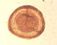
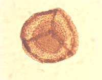
Spinozonotriletes uncatus Hacquebard, 1957
Radial, trilete, camerate miospores. Amb convexly rounded triangular. Trilete mark distinct, sutures straight though often obscured; extend to margin of inner body, accompanied by elevated flexuous folds of exoexine which extend decreasing in height, to the equatorial margin. Intexine thin, forms inner body, which is conformable in outline with the amb of the spore and occupies approximately three-quarters of the total spore diameter. Exoexine 3–6 µm thick, extended in the equatorial plane to form solid flange. Exoexine prominently ornamented with large spini which are concentrated in the equatorial region, commonly reduced in the distal polar region and completely absent from contact areas of proximal surface. Spini have broad, often bulbous bases and sharply pointed terminations; height 4–19 µm and basal diameter 2–6 µm. Spina size variable between specimens but uniform on any one specimen.
Hacquebard, 1957, p. 316, pl. 3, figs. 8–10. Playford, 1962, p. 657, pl. 94, figs. 4–6. Neves and Owens, 1966, pp. 355–356, pl. 3, figs. 2–3
Overall equatorial diameter excluding projecting ornament 74(104)150 µm
Spinozonotriletes conspicuus Playford 1964, and S. tenuispinosus Hacquebard 1957, are both similar but lack the elevated, flexuous, sutural folds present in S. uncatus.
Devonian to Mississippian; Tournaisian; LL to CM biozones (Higgs et al., 1988)
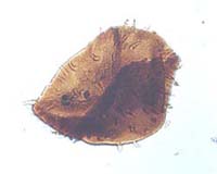
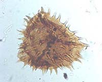
Torispora securis (Balme) Alpern, Doubinger & Horst, 1973
Monolete miospores. Amb ovoid, subtriangular or irregular. Miospore consists of ovoid to subcircular spore body with a pronounced patinate crassitude either over the distal surface or the distal and lateral surfaces. Monolete mark usually distinct, suture straight or curved; simple; length approximately three-quarters or more of the spore body radius. Suture commonly parallel to the long axis of the spore. Intexine of spore body relatively thin, laevigate. Exoexine thickened over part of the distal surface or at least in part in the equatorial plane to form patinate crassitude which may in some cases overlap a significant part of the spore body on the proximal surface. Profile of crassitude in compressed specimens crescentic or sub-rectangular, surface smooth or finely roughened. Boundary between crassitude and spore body gradational or abrupt.
Balme, 1952, p. 183. Smith and Butterworth, 1967, pp. 293–294, pl. 24, figs. 31–34
Overall equatorial diameter 26(34)44 µm. Diameter of inner body 15(21)28 µm
Smith and Butterworth (1967) did not differentiate Torispora securis, T. granulata Alpern 1959, T. laevigata Bharadwaj 1957 and T. verrucosa Alpern 1958.
Pennsylvanian; Moscovian to Gzhelian [Bolsovian to ?early Autunian]; SL to ?VC biozones (Clayton et al., 1977)
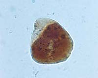
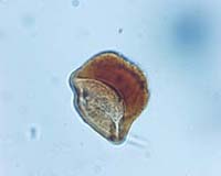
Tripartites trilinguis (Horst) Smith & Butterworth, 1967
Radial trilete miospores. Amb triangular, sides highly concave, apices rounded, giving trifoliate appearance. Trilete mark distinct, sutures simple, straight; extending half to two-thirds of spore radius. Exine extended laterally in the equatorial plane to form prominent rounded plicated or corrugated auriculae in the radial positions. Thickness of auriculae up to 6 µm, width 24–32 µm. Inter-radial areas deeply incised, narrow, occupied by markedly reduced inter-radial crassitude. Proximal exine laevigate but distal exine bears small well-spaced grana on the radial apices in an arc-shaped pattern between the ends of the trilete sutures and the inner margin of the auriculae. Exine of spore body thin.
Horst, 1955, p. 176, pl. 23, figs. 55–56. Smith and Butterworth, 1967, pp. 208–209. p1. 13, figs. 6–9
Overall equatorial diameter 32(44)51 µm
Tripartites trilinguis differs from T. vetustus in possessing more laterally expanded auriculae producing a markedly trifoliate appearance, and in possessing an arcuate distribution of grana between the ends of the trilete sutures and the inner margin of the auriculae. T. trilinguis also lacks the characteristic plications of the auriculae which characterise T. vetustus.
Mississippian; ?late Visean to early Serpukhovian [early Arnsbergian]; ?VF to TK biozones (Clayton et al., 1977, Owens et al., 1977)
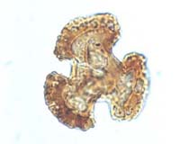
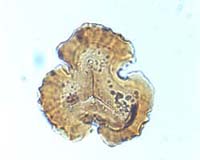
Tripartites vetustus Schemel, 1950
Radial, trilete, miospores. Amb triangular, sides slightly to strongly concave, apices rounded or truncated. Trilete mark distinct; sutures straight simple or accompanied by low narrow labra extending within a very short distance of the equatorial margin. Exine extended laterally in the equatorial plane to form well-developed auriculae in the radial positions, inter-radial connections between auriculae reduced or absent. Distal surface of auriculae plicated; remainder of exine laevigate.
Schemel, 1950, p. 243, pl. 40, fig. 11
Overall equatorial diameter 30(42)50 µm
Mississippian; late Visean to early Serpukhovian [early Arnsbergian], VF to TK biozones (Clayton et al., 1977, Owens et al., 1977)
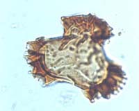
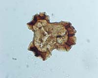
Triquitrites sculptilis (Balme) Smith & Butterworth, 1967
Radial, trilete, acamerate miospores. Amb irregularly triangular with more or less straight sides and rounded, flat or lobate apices. Shape dependent on the extent of exine ornamentation. Proximal surface flattened, distal surface inflated hemispherical. Trilete mark distinct to indistinct, sutures simple, straight; extending from two-thirds to the full spore radius. Proximal exine laevigate but distal exine ornamented with irregular pattern of ridges and verrucae. Verrucae variable in size and distribution; frequent fusion occurs between the bases of adjacent elements to produce an ill-defined reticulate pattern. Thickening of the exine at radial apices occurs to produce a variable crassitude. When associated with verrucae, this projects at the equatorial margin. Crassitudes, may in some cases, extend into inter-radial areas. Crassitudes vary from 2–7 µm across when measured along a given radius, and are 10–17 µm in width. Exine moderately thick.
Balme, 1952, p. 181, text-fig. 1g. Smith and Butterworth, 1967, p. 204, pl. 12, figs. 10–15
Overall equatorial diameter 25(37)45 µm
Smith and Butterworth (1967) regarded Triquitrites bucculentus Guennel 1958, and T. coesfeldens Bharadwaj 1957, as forms which fall within the extremes of the range of morphological variation displayed by T. sculptilis.
Pennsylvanian; Moscovian [late Duckmantian to early Asturian] (Smith and Butterworth, 1967)
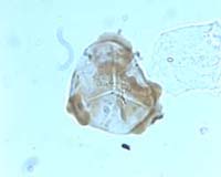
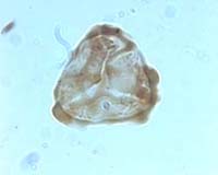
Triquitrites trivalvis (Waltz) Potonié & Kremp, 1956
Radial, trilete, acamerate miospores. Amb triangular to sub-triangular with rounded apices and straight to concave sides. Trilete mark distinct; sutures simple, straight, extend from two-thirds to the full radius of the spore body. Spore body more or less laevigate. Exoexine extended in the equatorial plane to produce variable cingulum which is narrow (2–4 µm) in the inter-radial areas but greatly expanded in the radial positions to form prominent auriculae. Outer margins of auriculae thickened and laterally expanded to create large, homogenous cushion-like caps with a smooth to crenulate surface. Caps approximately 7 µm across when measured along a radius, and are 30 µm wide.
Potonié and Kremp, 1956, p. 88. Playford, 1962, pp. 602–603, pl. 85, figs. 13–14
Overall equatorial diameter 38(51)66 µm. Diameter of spore body 24(34)44 µm
Mississippian; ?late Visean to early Serpukhovian [early Arnsbergian]; ?VF to TK biozones (Clayton et al., 1977, and Owens et al., 1977)
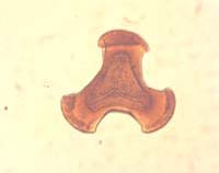
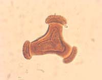
Umbonatisporites distinctus Clayton, 1971
Radial, trilete, acamerate miospores. Amb circular, subcircular or oval. Trilete mark often distinct; sutures when seen, simple, straight, extending between one-third and one-half the spore radius. Exine, 1–3 µm thick, ornamented on distal surface and equatorial portion of proximal surface with cylindrical shaft-like elements which are 2–10 µm high and 1–3 µm wide, with a prominent constriction near the top and a second near the base. Terminus of each shaft-like element normally flat but commonly surmounted by a single spina or conus up to 4.5 µm high. Rare small coni and spini may be interspersed between the longer biform elements. Ornamentation elements variable in size between specimens but commonly uniform on any single specimen. Contact areas laevigate.
Clayton, 1971, pp. 591–592, pl. 4, figs. 4–6
Overall equatorial diameter excluding ornamentation 43(70)126 µm
Umbonatisporites distinctus is distinguished from U. abstrusus (Playford) Clayton 1971 and U. variabilis Hibbert & Lacey 1969 by possessing cylindrical processes with flat or slightly concave tops surmounted by a single conus or spina, and by the uniformity of its ornamentation on any single specimen.
Mississippian; Tournaisian; HD to CM biozones (Higgs et al., 1988)
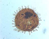
Umbonatisporites variabilis Hibbert & Lacey, 1969
Radial, trilete, acamerate miospores. Amb circular to subcircular. Trilete mark frequently indistinct or torn; sutures when seen are short, simple and straight, extending up to one-sixth of spore radius. Exine 1–2 µm thick, ornamented on distal surface and equatorial portion of the proximal surface with variable squat biform baculate or pilate projections arranged randomly over the surface. Elements variable in shape and size on any one specimen; individual elements up to 4.5 µm high and 1.0–1.5 µm in basal diameter, widening towards the apex. At the terminus of each element are one to three rounded heads, each surmounted by a single thin tapering spina. Between the larger biform elements, slender tapering spinae 2–4 µm long and up to 1 µm wide may occur.
Hibbert & Lacey, 1969, pp. 423–424, pl. 78, figs. 12–13, pl. 79, figs. 1–3
Overall equatorial diameter excluding projecting ornament 95(106)134 µm
Distinguished from Umbonatisporites distinctus Clayton 1971 by the multiple character of the spinose terminations surmounting the expanded baculate projections.
Mississippian; Visean to Serpukhovian [early Arnsbergian] (Hibbert and Lacey, 1969, and unpublished information)
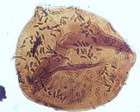
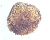
Vallatisporites agadesi Loboziak & Alpern, 1978
Radial, trilete, cingulizonate miospores. Amb sub-triangular to subcircular. Trilete mark distinct, sutures obscured by elevated flexuous folds of the exoexine which extend, decreasing in height, to the equatorial margin. Exine composed of two layers. Intexine forming inner body, thin with several concentric plications. Exoexine rugulate, completely enclosing inner body and extended in the equatorial plane to form a wide cingulizona containing a large number of small, radially orientated, internal vacuoles. Maximum thickness of cingulizona developed in the region adjacent to inner body. Distal surface of exoexine bears vermiculate to coarsely reticulate ornamentation. Ornamentation elements variable; irregular convolutions and rugulae common; verrucae and coni infrequent.
Loboziak & Alpern, 1978, pp. 58–59, pl. 2, figs. 9–12
Equatorial diameter 60(69)80 µm. Width of cingulizona 10(13)18 µm
Distinguished from Vallatisporites banffensis Staplin & Jansonius 1964, by the more coalescent nature of the ornament developed on the distal surface and the more obvious internal vacuolation of the zona. V. splendens Staplin & Jansonius 1964, differs by possessing a coarser ornament of more varied processes on the distal surface of the central body and less well-developed internal vacuolation of the cingulizona.
Mississippian; late Tournaisian to early Serpukhovian (Loboziak and Clayton, 1988)
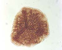
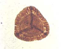
Vallatisporites drybrookensis Playford & McGregor, 1993
Radial, trilete, cingulizonate miospores. Amb rounded triangular with convex sides and rounded apices, sometimes subcircular. Trilete mark distinct; sutures straight, extending to the margin of the inner body, accompanied by elevated flexuous folds of the exoexine which extend, decreasing in height, to the equatorial margin. Intexine thin, forming rounded triangular inner body, frequently folded independently and withdrawn from the exoexine at least in the equatorial plane. Exoexine thin over proximal surface but thicker over distal surface and extended in the equatorial plane to form variably thickened cingulizona consisting of two zones. Inner thinner zone contains numerous internal radially orientated vacuoles separated by thin walls. Outer zone thicker and separated from the inner thinner zone by an irregular boundary coinciding with the limits of vacuolation. Distal surface of exoexine ornamented with pointed or rounded coni up to 3 µm high and 3.5 µm in basal diameter, interspersed with galeae consisting of a bulbous base surmounted by a small sharply pointed conus. Ornamentation reduced over the outer part of the cingulum to small coni. Well-developed cuniculus developed in the equatorial plane adjacent to the margin of the intexine.
Sullivan, 1964, pp. 370–371, pl. 59, figs. 14–15. Playford and McGregor, 1993, pp. 33–34, pl. 18, figs. 5, 6a,b, 7, 8a,b
Overall equatorial diameter 50(62)70 µm
Playford and McGregor (1993) considered that specimens assigned by Sullivan (1964) to his new combination Vallatisporites (Zonotriletes) ciliaris (Luber & Waltz) Sullivan 1964 were incorrectly identified. Consequently they erected Vallatisporites drybrookensis to include Sullivan’s specimens and designated a specimen of Sullivan’s as holotype.
The well-developed conate/galeate ornamentation and extensively internally vacuolate inner cingulizona distinguishes V. ciliaris from other species assigned to the genus Vallatisporites.
Mississippian to Pennsylvanian: early Tournaisian to ?Kazimovian/Gzhelian [Stephanian] (Playford and McGregor, 1993)
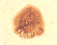
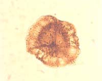
Vallatisporites hystricosus (Winslow) Byvsheva 1985
Radial, trilete, cinguli-camerate miospores. Amb rounded triangular to subcircular. Outline of inner body may be conformable with equatorial margin of spore or more circular resulting in the equatorial cinguli-zonate structure being wider in the radial positions. Trilete mark distinct, sutures straight, extending to margin of inner body, accompanied by elevated, flexuous folds of the exoexine, up to 4 µm in the polar region but decreasing in height and extending to the equatorial margin. Intexine thin, laevigate or finely infrapunctate, forming inner body. Exoexine closely adpressed to intexine except in the equatorial plane where a well developed cuniculus indicates limited separation. Exoexine extended in the equatorial plane to form bizonate cingulum consisting of a thinner, inner, extensively vacuolated zone with closely spaced radially orientated vacuoles separated by narrow walls, and a thicker, outer zone. Distal surface of exoexine bears galeate ornament of variable density; elements are largest and most densely distributed in the distal polar region and finer and less densely distributed towards the equatorial margin. Small, sharply pointed coni may be located in the spaces between the galeate elements. Individual galeae up to 10 µm high and 7 µm in basal diameter, consisting of a rounded basal boss surmounted by a sharply pointed, slender spina. Fusion between bases of adjacent elements forms irregular vermiculate elements with common spinose crests, particularly in the polar region. Numerous elements project at the equatorial margin.
Winslow, 1962, pp. 41–42, pl. 18, fig. 5
Overall equatorial diameter 46–84 µm
Vallatisporites pusillites (Kedo) Dolby & Neves 1970 is similar in many respects to Cirratriradites hystricosus but is distinguished by possessing relatively finer ornamentation in which non-galeate elements are more common. It is probable that complete morphological gradation exists between the two species. Vallatisporites verrucosus Hacquebard 1957 is distinguished by possessing a verrucose ornament. Vallatisporites galearis Sullivan 1964 is smaller and possesses an ornament of galeae with cone-shaped rather than long, spinose terminations.
Late Devonian to Mississippian; Strunian to Tournaisian (Winslow, 1962)
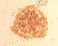
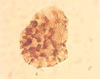
Vallatisporites vallatus Hacquebard, 1957
Radial, trilete, camerate, cingulizonate miospores. Amb convexly rounded triangular. Trilete mark distinct to indistinct, sutures straight, extending to margin of inner body, accompanied by low, slightly elevated exoexinal folds which decrease in height towards the ends of the sutures. Height of folds in polar region approximately 2.5 µm. Exine two layered. Intexine thin, forms inner body conformable in outline with equatorial margin, surface laevigate. Exoexine closely adpressed to intexine over proximal and distal surface but separated at least to some extent in the equatorial plane where a well-developed narrow cuniculus is developed. Exoexine thin over proximal surface but slightly thicker distally and extended in the equatorial plane to form differentially thickened tapering cingulum. Cingulum consists of inner thicker zone that appears in part to overlap the equatorial margin of the inner body, up to 8 µm wide. In many specimens large numbers of small elongate internal vacuoles, separated by narrow walls, appear to be located within this portion of the cingulum. Individual vacuoles up to 5 µm in length. Outer part of cingulum thin and sometimes weakly radially striated. Distal exoexine bears a dense ornament of coni, grana and small spini, elements up to 3 µm in height and 2 µm in basal diameter. Elements normally discrete but some basal coalescence occurs. Ornament more sparsely distributed on the cingulum and at the equatorial margin is limited to small conate projections.
Hacquebard, 1957, pp. 312–313, pl. 2, fig. 12. Staplin and Jansonius, 1964, p. 112, p1. 21, figs. 1–6. Higgs, Clayton and Keegan, 1988, pp. 80–81, pl. 16, figs. 19–20
Overall equatorial diameter 50(59)70 µm. Diameter of spore body 35–44 µm
Vallatisporites vallatus is distinguished from V. verrucosus Hacquebard 1957 by possessing a fine densely distributed ornament of coni, grana and infrequent spini that are less than 3 µm in height. V. pusillites (Kedo) Dolby & Neves 1960, differs by possessing an ornament of coarser coni and galeae 4–12 µm high with bases up to 6 µm in diameter.
Late Devonian to Mississippian; Tournaisian, VI to CM biozones (Higgs et al., 1988)
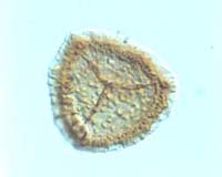
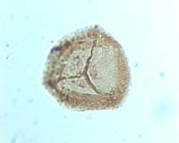
Vallatisporites verrucosus Hacquebard, 1957
Radial, trilete, camerate, cingulizonate miospores. Amb convexly rounded triangular. Trilete mark distinct or indistinct if obscured by ornament; sutures straight, extending to margin of inner body, simple or accompanied by elevated flexuous folds of the exoexine, up to 4 µm high in polar region, which extend, decreasing in height, to the margin of the body or onto the cingulum. Exine two layered. Intexine thin, laevigate, forms inner body conformable with the equatorial outline. Exoexine closely adpressed to intexine in proximal and distal positions but separated from intexine in the equatorial plane where a well-developed cuniculus is located. Exoexine extended in the equatorial plane to form poorly differentiated bizonate cingulum. Inner thicker zone of cingulum overlaps the equatorial margin of inner body and may contain numerous internal radially orientated vacuoles. Outer zone of cingulum thinner and narrower. In some specimens differentiation of the cingulum is difficult to discern. Distal surface of exoexine densely ornamented with discrete or confluent verrucae up to 6 µm in diameter. Elements may fuse to form short irregular ridges. Verrucae are most densely distributed in the distal polar region where the largest elements are also located. Towards the equatorial margin, elements become progressively smaller and more sparsely distributed. Equatorial margin uneven but rarely displays coarse projecting ornamentation. Verrucae surmounted by small mammoid coni may be present in the distal polar region.
Hacquebard, 1957, p. 313, pl. 2, fig. 13
Overall equatorial diameter 55–75 µm. Diameter of inner body 41–47 µm
Almost identical in structure to Vallatisporites vallatus Hacquebard 1957, but distinguished by possessing an ornament of verrucate elements and by the poorly differentiated bizonate cingulum.
Late Devonian to Mississipian; Strunian to Tournaisian; LN to CM biozones (Higgs et al., 1988)